Explore
 Validate
 Learn
Learn Western blot
Antibody data
- Antibody Data
- Antigen structure
- References [1]
- Comments [0]
- Validations
- Western blot [1]
- Immunohistochemistry [1]
- Flow cytometry [6]
- Other assay [1]
Submit
Validation data
Reference
Comment
Report error
- Product number
- MA1-41019 - Provider product page

- Provider
- Invitrogen Antibodies
- Product name
- CD254 (RANK Ligand) Monoclonal Antibody (12A380)
- Antibody type
- Monoclonal
- Antigen
- Other
- Description
- Suggested positive control: activated T cells or RAW cells, antigen standard for TNFSF11 (transient overexpression lysate).
- Reactivity
- Human, Mouse
- Host
- Mouse
- Isotype
- IgG
- Antibody clone number
- 12A380
- Vial size
- 100 μg
- Concentration
- 1.0 mg/mL
- Storage
- Store at 4°C short term. For long term storage, store at -20°C, avoiding freeze/thaw cycles.
Submitted references Bu‑Shen‑Ning‑Xin decoction suppresses osteoclastogenesis by modulating RANKL/OPG imbalance in the CD4+ T lymphocytes of ovariectomized mice.
Zhang JL, Qiu XM, Zhang N, Tang W, Gober HJ, Li DJ, Wang L
International journal of molecular medicine 2018 Jul;42(1):299-308
International journal of molecular medicine 2018 Jul;42(1):299-308
No comments: Submit comment
Supportive validation
- Submitted by
- Invitrogen Antibodies (provider)
- Main image
- Experimental details
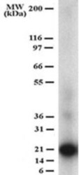
- Western blot analysis of CD254 (RANK Ligand) in recombinant RANKL (partial). Samples were incubated in CD254 (RANK Ligand) monoclonal antibody (Product # MA1-41019) using a dilution of 1 µg/mL.

Supportive validation
- Submitted by
- Invitrogen Antibodies (provider)
- Main image

- Experimental details
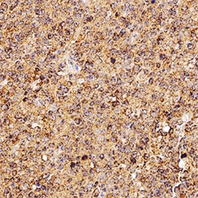
- Immunohistochemical analysis of CD254 (RANK Ligand) in formalin-fixed paraffin-embedded human tonsil. Samples were incubated in CD254 (RANK Ligand) monoclonal antibody (Product # MA1-41019) using a dilution of 0.0763888888888889. Bond Rx autostainer (Leica Biosystems). The assay involved 20 minutes of heat induced antigen retrieval (HIER) using 10mM sodium citrate buffer (pH 6.0) and endogenous peroxidase quenching with peroxide block. The sections were incubated with primary antibody for 30 minutes and Bond Polymer Refine Detection (Leica Biosystems) with DAB was used for signal development followed by counterstaining with hematoxylin. Whole slide scanning and capturing of representative images was performed using Aperio AT2 (Leica Biosystems). Cytoplasmic staining was observed. Staining was performed by Histowiz.
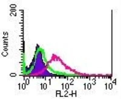
Supportive validation
- Submitted by
- Invitrogen Antibodies (provider)
- Main image

- Experimental details
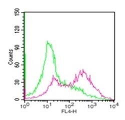
- Flow cytometry of of CD254 (RANK Ligand) in RAW cells. Frozen samples were incubated in CD254 (RANK Ligand) monoclonal antibody (Product # MA1-41019) using a dilution of 2 µg/mL. Shaded histogram represents cells without antibody; green represents isotype control antibody; purple represents antibody. Image using the PE format of this antibody.

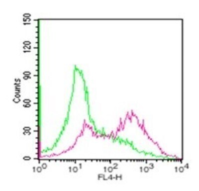
- Submitted by
- Invitrogen Antibodies (provider)
- Main image
- Experimental details
- Flow cytometry of CD254 (RANK Ligand) in RANKL in untreated mouse splenocytes (green) and 72 hour ConA-stimulated mouse splenocytes (red). Samples were incubated in CD254 (RANK Ligand) monoclonal antibody (Product # MA1-41019) using a dilution of 1.0 µg. Antibody was conjugated to Alexa Fluor (R) 647.

- Submitted by
- Invitrogen Antibodies (provider)
- Main image

- Experimental details
- Flow cytometry of CD254 (RANK Ligand) in RANKL in untreated mouse splenocytes (green) and 72 hour ConA-stimulated mouse splenocytes (red). Samples were incubated in CD254 (RANK Ligand) monoclonal antibody (Product # MA1-41019) using a dilution of 1.0 µg. Antibody was conjugated to Alexa Fluor (R) 488.

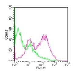
- Submitted by
- Invitrogen Antibodies (provider)
- Main image

- Experimental details
- Flow cytometry of of CD254 (RANK Ligand) in RAW cells. Frozen samples were incubated in CD254 (RANK Ligand) monoclonal antibody (Product # MA1-41019) using a dilution of 2 µg/mL. Shaded histogram represents cells without antibody; green represents isotype control antibody; purple represents antibody. Image using the PE format of this antibody.
- Submitted by
- Invitrogen Antibodies (provider)
- Main image

- Experimental details
- Flow cytometry of CD254 (RANK Ligand) in RANKL in untreated mouse splenocytes (green) and 72 hour ConA-stimulated mouse splenocytes (red). Samples were incubated in CD254 (RANK Ligand) monoclonal antibody (Product # MA1-41019) using a dilution of 1.0 µg. Antibody was conjugated to Alexa Fluor (R) 647.
- Submitted by
- Invitrogen Antibodies (provider)
- Main image
- Experimental details
- Flow cytometry of CD254 (RANK Ligand) in RANKL in untreated mouse splenocytes (green) and 72 hour ConA-stimulated mouse splenocytes (red). Samples were incubated in CD254 (RANK Ligand) monoclonal antibody (Product # MA1-41019) using a dilution of 1.0 µg. Antibody was conjugated to Alexa Fluor (R) 488.

Supportive validation
- Submitted by
- Invitrogen Antibodies (provider)
- Main image

- Experimental details
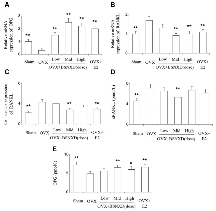
- Figure 3 Modulatory effect of BSNXD on RANKL and OPG. Following 8 weeks of BSNXD administration, mice in six experimental groups were sacrificed, and single cells were isolated from the spleen by means of density gradient centrifugation. CD4 + T cells were isolated from single cells via magnetic bead selection. The CD4 + T cells were separated into three portions for testing. One portion was used to estimate the relative mRNA expression of RANKL and OPG by reverse transcription-quantitative polymerase chain reaction, another was used to determine the cell surface expression of RANKL by flow cytometry, and the third was cultured in vitro and the supernatant harvested to detect the sRANKL and OPG levels by ELISA. (A) Relative mRNA expression of RANKL, (B) relative mRNA expression of OPG and (C) cell surface expression of RANKL. Protein levels of (D) RANKL and (E) OPG in the cell culture supernatant. Data are expressed as the mean +- standard error of the mean. ** P