Explore
 Validate
 Learn
LearnNBP1-30032
antibody from Novus Biologicals
Targeting: ABCA4
ABCR, ARMD2, CORD3, FFM, RP19, STGD, STGD1
 Western blot
 Immunocytochemistry
Antibody data
- Antibody Data
- Antigen structure
- References [4]
- Comments [0]
- Validations
- Western blot [1]
- Immunohistochemistry [4]
Submit
Validation data
Reference
Comment
Report error
- Product number
- NBP1-30032 - Provider product page

- Provider
- Novus Biologicals
- Proper citation
- Novus Cat#NBP1-30032, RRID:AB_1968439
- Product name
- Mouse Monoclonal ABCA4 Antibody
- Antibody type
- Monoclonal
- Description
- Protein G purified.
- Reactivity
- Human, Mouse, Bovine, Canine, Xenopus
- Host
- Mouse
- Isotype
- IgG
- Vial size
- 0.1 ml
- Storage
- Store at -20C. Avoid freeze-thaw cycles.
Submitted references An ABCA4 loss-of-function mutation causes a canine form of Stargardt disease.
ABCA4 mutations causing mislocalization are found frequently in patients with severe retinal dystrophies.
The 220-kDa rim protein of retinal rod outer segments is a member of the ABC transporter superfamily.
The 220-kDa rim protein of retinal rod outer segments is a member of the ABC transporter superfamily.
Mäkeläinen S, Gòdia M, Hellsand M, Viluma A, Hahn D, Makdoumi K, Zeiss CJ, Mellersh C, Ricketts SL, Narfström K, Hallböök F, Ekesten B, Andersson G, Bergström TF
PLoS genetics 2019 Mar;15(3):e1007873
PLoS genetics 2019 Mar;15(3):e1007873
ABCA4 mutations causing mislocalization are found frequently in patients with severe retinal dystrophies.
Wiszniewski W, Zaremba CM, Yatsenko AN, Jamrich M, Wensel TG, Lewis RA, Lupski JR
Human molecular genetics 2005 Oct 1;14(19):2769-78
Human molecular genetics 2005 Oct 1;14(19):2769-78
The 220-kDa rim protein of retinal rod outer segments is a member of the ABC transporter superfamily.
Illing M, Molday LL, Molday RS
The Journal of biological chemistry 1997 Apr 11;272(15):10303-10
The Journal of biological chemistry 1997 Apr 11;272(15):10303-10
The 220-kDa rim protein of retinal rod outer segments is a member of the ABC transporter superfamily.
Illing M, Molday LL, Molday RS
The Journal of biological chemistry 1997 Apr 11;272(15):10303-10
The Journal of biological chemistry 1997 Apr 11;272(15):10303-10
No comments: Submit comment
Supportive validation
- Submitted by
- Novus Biologicals (provider)
- Main image
- Experimental details
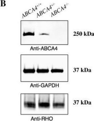
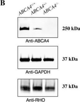
- Western Blot: ABCA4 Antibody (3F4) [NBP1-30032] - Characterization of ABCA4 mRNA expression and western blot analyses of ABCA4 protein levels in the canine retina. Western blot analyses of ABCA4 (above), GAPDH (middle), and RHO (below) protein levels in retinal tissue of dogs with the three different genotypes. Image collected and cropped by CiteAb from the following publication (http://dx.plos.org/10.1371/journal.pgen.1007873), licensed under a CC-BY licence.
Supportive validation
- Submitted by
- Novus Biologicals (provider)
- Main image
- Experimental details
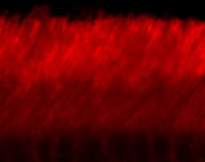
- Immunohistochemistry: ABCA4 Antibody (3F4) [NBP1-30032] - staining of adult mouse retina showing specific immunolabeling of the ABCA4 protein. Photo courtesy of Mary Raven, University of California, Santa Barbara, CA.
- Submitted by
- Novus Biologicals (provider)
- Main image
- Experimental details
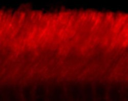
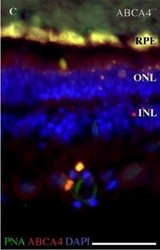
- Immunohistochemistry: ABCA4 Antibody (3F4) [NBP1-30032] - Fluorescence histochemistry of ABCA4, cone photoreceptors, and autofluorescence in the canine retina. Fluorescence micrographs showing ABCA4 expression (red), FITC-conjugated peanut agglutinin (PNA, green), and DAPI nuclear staining (blue) in affected (ABCA4-/-) retina. PNA labels cone photoreceptors. Autofluorescence, indicative of lipofuscin accumulation, was seen in the ABCA4-/- RPE. Image collected and cropped by CiteAb from the following publication (http://dx.plos.org/10.1371/journal.pgen.1007873), licensed under a CC-BY licence.

- Submitted by
- Novus Biologicals (provider)
- Main image
- Experimental details
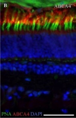
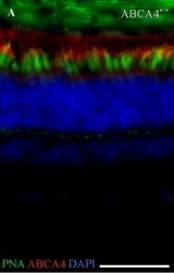
- Immunohistochemistry: ABCA4 Antibody (3F4) [NBP1-30032] - Fluorescence histochemistry of ABCA4, cone photoreceptors, and autofluorescence in the canine retina. Fluorescence micrographs showing ABCA4 expression (red), FITC-conjugated peanut agglutinin (PNA, green), and DAPI nuclear staining (blue) in wild-type (ABCA4+/+) retina. PNA labels cone photoreceptors. Image collected and cropped by CiteAb from the following publication (http://dx.plos.org/10.1371/journal.pgen.1007873), licensed under a CC-BY licence.

- Submitted by
- Novus Biologicals (provider)
- Main image
- Experimental details
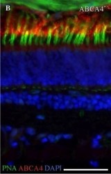
- Immunohistochemistry: ABCA4 Antibody (3F4) [NBP1-30032] - Fluorescence histochemistry of ABCA4, cone photoreceptors, and autofluorescence in the canine retina. Fluorescence micrographs showing ABCA4 expression (red), FITC-conjugated peanut agglutinin (PNA, green), and DAPI nuclear staining (blue) in heterozygous (ABCA4+/-) retina. PNA labels cone photoreceptors. Image collected and cropped by CiteAb from the following publication (http://dx.plos.org/10.1371/journal.pgen.1007873), licensed under a CC-BY licence.