Explore
 Validate
 Learn
Learn Western blot
 Immunocytochemistry
Antibody data
- Antibody Data
- Antigen structure
- References [0]
- Comments [0]
- Validations
- Immunocytochemistry [1]
- Immunoprecipitation [2]
- Protein array [2]
- Chromatin Immunoprecipitation [1]
Submit
Validation data
Reference
Comment
Report error
- Product number
- MA1-2021 - Provider product page

- Provider
- Invitrogen Antibodies
- Product name
- Anti-Acetyl Lysine
- Antibody type
- Monoclonal
- Antigen
- Synthetic peptide sequence surrounding the acetylated lysine 9 of histone H3.
- Reactivity
- Human, Mouse
- Host
- Mouse
- Isotype
- IgG
- Antibody clone number
- 1C6
- Vial size
- 100 µg
- Concentration
- 1 mg/ml
- Storage
- -20°C, Avoid Freeze/Thaw Cycles
No comments: Submit comment
Supportive validation
- Submitted by
- Invitrogen Antibodies (provider)
- Main image
- Experimental details
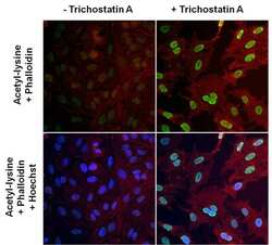
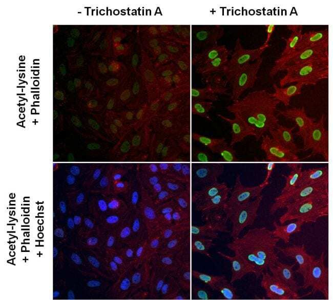
- Immunofluorescent analysis of lysine acetylated proteins (green) in HeLa cells left untreated (left panels) or treated with 0.3uM Trichostatin A for 16 hours (right panels). Formalin fixed cells were permeabilized with 0.1% Triton X-100 in TBS for 10 minutes at room temperature and blocked with 1% Blocker BSA (Product # 37525) for 15 minutes at room temperature. Cells were probed with an Acetyl Lysine monoclonal antibody (Product # MA1-2021) at a dilution of 1:100 for 1 hour at room temperature, washed with PBS, and incubated with DyLight 488 goat anti-mouse IgG secondary antibody (Product # 35502) at a dilution of 1:400 for 30 minutes at room temperature. F-Actin (red) was stained with DyLight 554 Phalloidin (Product # 21834). A merged image with nuclei staining (blue) using Hoechst 33342 dye (Product # 62249) is shown on bottom panels. Images were taken on a Thermo Scientific ArrayScan at 20X magnification.
Supportive validation
- Submitted by
- Invitrogen Antibodies (provider)
- Main image
- Experimental details
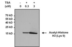
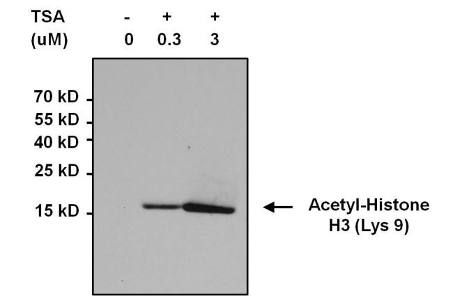
- Immunoprecipitation of acetylated Histone H3 (Lys9) was performed using whole cell lysates from cells left untreated (DMSO only) or cells treated with 0.3uM or 3uM Trichostatin A (TSA) for 16 hours. Antigen-antibody complexes were formed by incubating 500 µg of the indicated lysate with 3 µg of an Acetyl Lysine monoclonal antibody (Product # MA1-2021) overnight on a rocking platform at 4°C. The immune complexes were captured on 50 µL Protein A/G Agarose (Product # 20421), washed extensively, and eluted with 5X Lane Marker Reducing Sample Buffer (Product # 39000). Samples were resolved on a 4-20% Tris-HCl polyacrylamide gel, transferred to a PVDF membrane, and blocked with 5% BSA/TBS-0.1%Tween for at least 1 hour. The membrane was probed with an Acetyl-Histone H3 (Lys9) monoclonal antibody (Product # MA5-11195) at a dilution of 1:1000 overnight rotating at 4°C, washed in TBST, and probed with Clean-blot IP Detection Reagent (Product # 21230) at a dilution of 1:2000 for at least 1 hour. Chemiluminescent detection was performed using SuperSignal West Pico (Product # 34087).
- Submitted by
- Invitrogen Antibodies (provider)
- Main image

- Experimental details
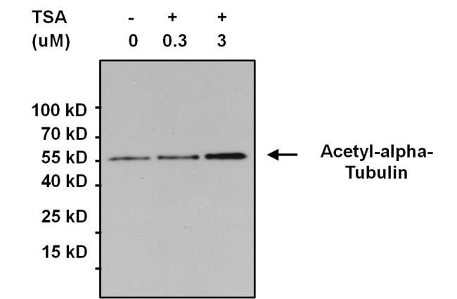
- Immunoprecipitation of acetylated alpha-Tubulin was performed using whole cell lysates from cells left untreated (DMSO only) or cells treated with 0.3uM or 3uM Trichostatin A (TSA) for 16 hours. Antigen-antibody complexes were formed by incubating 500 µg of lysate with 3 µg of an Acetyl Lysine monoclonal antibody (Product # MA1-2021) overnight on a rocking platform at 4°C. The immune complexes were captured on 50 µL Protein A/G Agarose (Product # 20421), washed extensively, and eluted with 5X Lane Marker Reducing Sample Buffer (Product # 39000). Samples were resolved on a 4-20% Tris-HCl polyacrylamide gel, transferred to a PVDF membrane, and blocked with 5% BSA/TBS-0.1%Tween for at least 1 hour. The membrane was probed with an alpha-Tubulin polyclonal antibody (Product # PA5-19489) at a dilution of 1:1000 overnight rotating at 4°C, washed in TBST, and probed with Clean-blot IP Detection Reagent (Product # 21230) at a dilution of 1:2000 for at least 1 hour. Chemiluminescent detection was performed using SuperSignal West Pico (Product # 34087).
Supportive validation
- Submitted by
- Invitrogen Antibodies (provider)
- Main image
- Experimental details
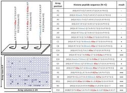
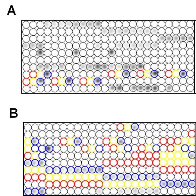
- Specific detection of lysine acetylated histones using an Acetyl Lysine monoclonal antibody (Product # MA1-2021). Histone peptides containing 384 different modification combinations spotted in duplicate on a peptide array were used according to the manufacturers instructions (Active Motif). A schematic and table with representative results (positive detection in red, negative in blue) are shown. Result interpretation and quantification were performed using the manufacturers software. Chemiluminescent detection was performed using SuperSignal West Pico (Product # 34087). Images were captured using a Thermo Scientific myECL Imager (Product # 62236).
- Submitted by
- Invitrogen Antibodies (provider)
- Main image
- Experimental details
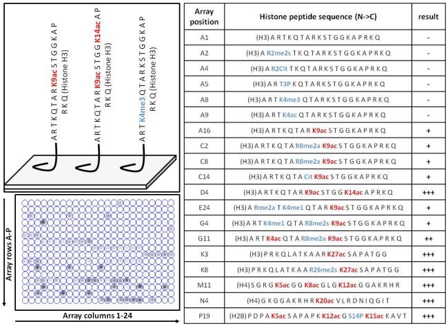
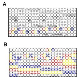
- Specific detection of lysine acetylated histones using an Acetyl Lysine monoclonal antibody (Product # MA1-2021). Histone peptides containing 384 different modification combinations spotted in duplicate on a peptide array were used according to the manufacturers instructions (Active Motif). Representative results showing specific detection of acetylated Histone H4 K20 (blue circles, panel A) and H3 K9 (blue circles, panel B). Note that Histone H4 K20me2/K20me3 (red and yellow circles, panel A) and H3 K9me2/K9m3 (red and yellow circles, panel B) are not detected by the Acetyl Lysine monoclonal antibody (Product # MA1-2021). Result interpretation and quantification were performed using the manufacturers software. Chemiluminescent detection was performed using SuperSignal West Pico (Product # 34087). Images were captured using a Thermo Scientific myECL Imager (Product # 62236).
Supportive validation
- Submitted by
- Invitrogen Antibodies (provider)
- Main image
- Experimental details
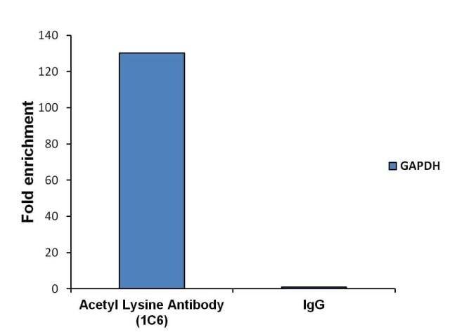
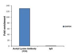
- Chromatin immunoprecipitation analysis (ChIP) of chromatin proteins with acetylated lysines was performed using cross-linked chromatin from 4x10^6 LNCaP cells. Immunoprecipitation was performed using the Pierce Magnetic ChIP kit (Product # 26157) with 10 µg of an Acetyl Lysine monoclonal antibody (Product # MA1-2021). Quantitative real-time PCR data was obtained using a Thermo PikoReal PCR System (Product # TCR0096), SYBR Green PCR Master Mix, and primers (5-TACTAGCGGTTTTACGGGCG, 3-TCGAACAGGAGGAGCAGAGAGCGA) flanking the human GAPDH promoter proximal to the start site of transcription. Quantitation of immunoprecipitated GAPDH promoter sequence is presented as fold enrichment of the acetyl lysine monoclonal antibody versus non-specific IgG.