Explore
 Validate
 Learn
Learn Western blot
Antibody data
- Antibody Data
- Antigen structure
- References [4]
- Comments [0]
- Validations
- Western blot [1]
- Immunocytochemistry [1]
Submit
Validation data
Reference
Comment
Report error
- Product number
- AP2402a - Provider product page

- Provider
- Abcepta
- Proper citation
- Abgent Cat#AP2402a, RRID:AB_2224813
- Product name
- AGL Antibody (Center)
- Antibody type
- Polyclonal
- Antigen
- Synthetic peptide
- Description
- Purified Rabbit Polyclonal Antibody (Pab)
- Reactivity
- Human
- Host
- Rabbit
- Isotype
- IgG
- Vial size
- 400 µl
- Concentration
- 2 mg/ml
- Storage
- Maintain refrigerated at 2-8°C for up to 6 months. For long term storage store at -20°C in small aliquots to prevent freeze-thaw cycles.
Submitted references Genetic depletion of the malin E3 ubiquitin ligase in mice leads to lafora bodies and the accumulation of insoluble laforin.
Fast-twitch sarcomeric and glycolytic enzyme protein loss in inclusion body myositis.
Abnormal metabolism of glycogen phosphate as a cause for Lafora disease.
A role for AGL ubiquitination in the glycogen storage disorders of Lafora and Cori's disease.
DePaoli-Roach AA, Tagliabracci VS, Segvich DM, Meyer CM, Irimia JM, Roach PJ
The Journal of biological chemistry 2010 Aug 13;285(33):25372-81
The Journal of biological chemistry 2010 Aug 13;285(33):25372-81
Fast-twitch sarcomeric and glycolytic enzyme protein loss in inclusion body myositis.
Parker KC, Kong SW, Walsh RJ, Bch, Salajegheh M, Moghadaszadeh B, Amato AA, Nazareno R, Lin YY, Krastins B, Sarracino DA, Beggs AH, Pinkus JL, Greenberg SA
Muscle & nerve 2009 Jun;39(6):739-53
Muscle & nerve 2009 Jun;39(6):739-53
Abnormal metabolism of glycogen phosphate as a cause for Lafora disease.
Tagliabracci VS, Girard JM, Segvich D, Meyer C, Turnbull J, Zhao X, Minassian BA, Depaoli-Roach AA, Roach PJ
The Journal of biological chemistry 2008 Dec 5;283(49):33816-25
The Journal of biological chemistry 2008 Dec 5;283(49):33816-25
A role for AGL ubiquitination in the glycogen storage disorders of Lafora and Cori's disease.
Cheng A, Zhang M, Gentry MS, Worby CA, Dixon JE, Saltiel AR
Genes & development 2007 Oct 1;21(19):2399-409
Genes & development 2007 Oct 1;21(19):2399-409
No comments: Submit comment
Supportive validation
- Submitted by
- Abcepta (provider)
- Main image

- Experimental details
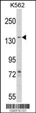
- Western blot analysis of hAGL-C371 (Cat. #AP2402a) in K562 cell line lysates (35ug/lane). AGL (arrow) was detected using the purified Pab.
- Primary Ab dilution
- 1:1000
Supportive validation
- Submitted by
- Abcepta (provider)
- Main image

- Experimental details
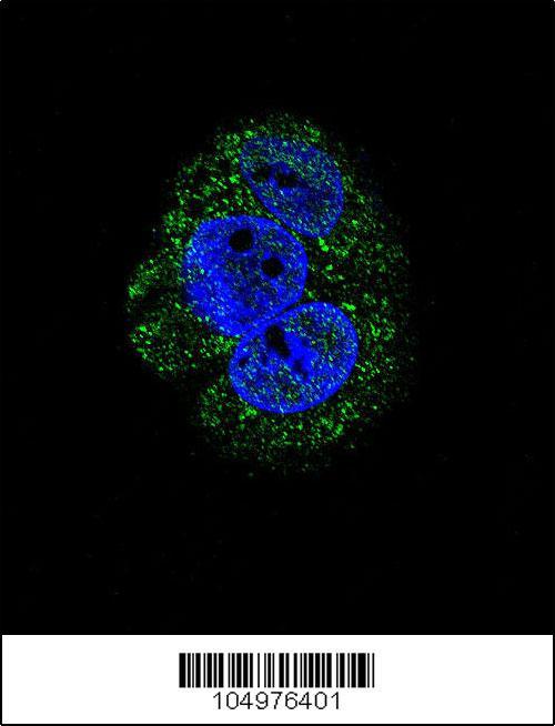
- Confocal immunofluorescent analysis of AGL Antibody (Center)(Cat#AP2402a) with HepG2 cell followed by Alexa Fluor 488-conjugated goat anti-rabbit lgG (green). DAPI was used to stain the cell nuclear (blue).
- Primary Ab dilution
- 1:10~50