Explore
 Validate
 Learn
Learn Western blot
Antibody data
- Antibody Data
- Antigen structure
- References [5]
- Comments [0]
- Validations
- Western blot [1]
- Immunocytochemistry [2]
Submit
Validation data
Reference
Comment
Report error
- Product number
- AP2402B - Provider product page

- Provider
- Abcepta
- Proper citation
- Abgent Cat#AP2402b, RRID:AB_2305340
- Product name
- AGL Antibody (C-term)
- Antibody type
- Polyclonal
- Antigen
- Synthetic peptide
- Description
- Purified Rabbit Polyclonal Antibody (Pab)
- Reactivity
- Human
- Host
- Rabbit
- Isotype
- IgG
- Vial size
- 400 µl
- Concentration
- 2 mg/ml
- Storage
- Maintain refrigerated at 2-8°C for up to 6 months. For long term storage store at -20°C in small aliquots to prevent freeze-thaw cycles.
Submitted references Laforin-malin complex degrades polyglucosan bodies in concert with glycogen debranching enzyme and brain isoform glycogen phosphorylase.
Anti-retinal antibodies in patients with macular telangiectasia type 2.
Genetic depletion of the malin E3 ubiquitin ligase in mice leads to lafora bodies and the accumulation of insoluble laforin.
Fast-twitch sarcomeric and glycolytic enzyme protein loss in inclusion body myositis.
A role for AGL ubiquitination in the glycogen storage disorders of Lafora and Cori's disease.
Liu Y, Zeng L, Ma K, Baba O, Zheng P, Liu Y, Wang Y
Molecular neurobiology 2014 Apr;49(2):645-57
Molecular neurobiology 2014 Apr;49(2):645-57
Anti-retinal antibodies in patients with macular telangiectasia type 2.
Zhu L, Shen W, Zhu M, Coorey NJ, Nguyen AP, Barthelmes D, Gillies MC
Investigative ophthalmology & visual science 2013 Aug 21;54(8):5675-83
Investigative ophthalmology & visual science 2013 Aug 21;54(8):5675-83
Genetic depletion of the malin E3 ubiquitin ligase in mice leads to lafora bodies and the accumulation of insoluble laforin.
DePaoli-Roach AA, Tagliabracci VS, Segvich DM, Meyer CM, Irimia JM, Roach PJ
The Journal of biological chemistry 2010 Aug 13;285(33):25372-81
The Journal of biological chemistry 2010 Aug 13;285(33):25372-81
Fast-twitch sarcomeric and glycolytic enzyme protein loss in inclusion body myositis.
Parker KC, Kong SW, Walsh RJ, Bch, Salajegheh M, Moghadaszadeh B, Amato AA, Nazareno R, Lin YY, Krastins B, Sarracino DA, Beggs AH, Pinkus JL, Greenberg SA
Muscle & nerve 2009 Jun;39(6):739-53
Muscle & nerve 2009 Jun;39(6):739-53
A role for AGL ubiquitination in the glycogen storage disorders of Lafora and Cori's disease.
Cheng A, Zhang M, Gentry MS, Worby CA, Dixon JE, Saltiel AR
Genes & development 2007 Oct 1;21(19):2399-409
Genes & development 2007 Oct 1;21(19):2399-409
No comments: Submit comment
Supportive validation
- Submitted by
- Abcepta (provider)
- Main image
- Experimental details
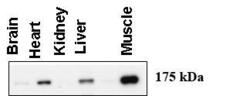
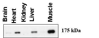
- "Western blot using anti-AGL (C-term) antibody (AP2402c) at 1:1000 dilution. A total of 20 ug of lysates was loaded for each tissue. Data courtesy of Dr. Alan Cheng, Department of Internal Medicine, Life Sciences Institute, University of Michigan Medical Center, Ann Arbor, Michigan."
- Primary Ab dilution
- 1:1000
Supportive validation
- Submitted by
- Abcepta (provider)
- Main image
- Experimental details
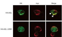
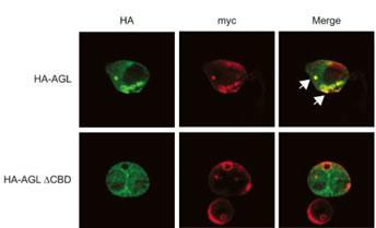
- Expression of myc-GS causes wild type but not the ÃCBD mutant of AGL to aggregate around the PAS-stain-positive inclusions. HepG2 cells were transfected with either HA-tagged wild-type AGL (HA-AGL) or HA-AGL ÃCBD. Cells were fixed in formalin and processed for IF using anti-HA (green) and anti-myc (red) antibodies. White arrows indicate colocalization of HA-AGL and myc-GS.
- Primary Ab dilution
- 1:10~50
- Submitted by
- Abcepta (provider)
- Main image

- Experimental details
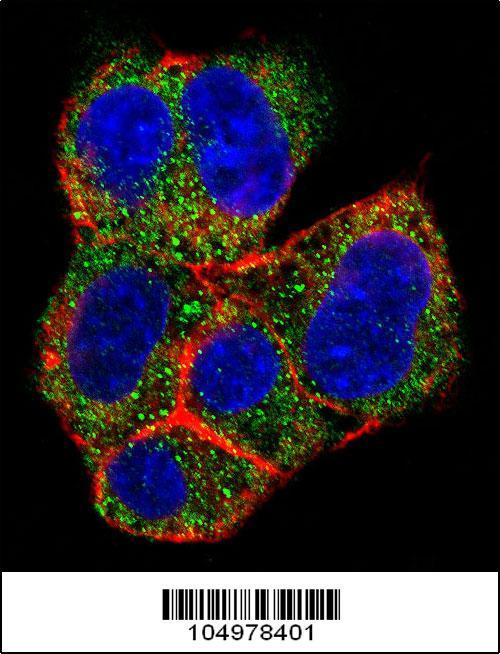
- Confocal immunofluorescent analysis of AGL Antibody (C-term)(Cat#AP2402b) with HepG2 cell followed by Alexa Fluor 488-conjugated goat anti-rabbit lgG (green). Actin filaments have been labeled with Alexa Fluor 555 phalloidin (red).DAPI was used to stain the cell nuclear (blue).
- Primary Ab dilution
- 1:10~50