Explore
 Validate
 Learn
Learn Immunocytochemistry
Antibody data
- Antibody Data
- Antigen structure
- References [1]
- Comments [0]
- Validations
- Immunocytochemistry [1]
- Other assay [1]
Submit
Validation data
Reference
Comment
Report error
- Product number
- PA5-66855 - Provider product page

- Provider
- Invitrogen Antibodies
- Product name
- PKP4 Polyclonal Antibody
- Antibody type
- Polyclonal
- Antigen
- Recombinant protein fragment
- Description
- Immunogen sequence: SYSDSGYQEAG SFHNSQNVSK ADNRQQHSFI GSTNNHVVRN SRAEGQTLVQ PSVANRAMRR VSSVPSRAQS PSYVISTGVS PSR Highest antigen sequence identity to the following orthologs - mouse 89%, rat 90%.
- Reactivity
- Human
- Host
- Rabbit
- Isotype
- IgG
- Vial size
- 100 μL
- Concentration
- 0.3 mg/mL
- Storage
- Store at 4°C short term. For long term storage, store at -20°C, avoiding freeze/thaw cycles.
Submitted references Kir2.1 Interactome Mapping Uncovers PKP4 as a Modulator of the Kir2.1-Regulated Inward Rectifier Potassium Currents.
Park SS, Ponce-Balbuena D, Kuick R, Guerrero-Serna G, Yoon J, Mellacheruvu D, Conlon KP, Basrur V, Nesvizhskii AI, Jalife J, Rual JF
Molecular & cellular proteomics : MCP 2020 Sep;19(9):1436-1449
Molecular & cellular proteomics : MCP 2020 Sep;19(9):1436-1449
No comments: Submit comment
Supportive validation
- Submitted by
- Invitrogen Antibodies (provider)
- Main image
- Experimental details
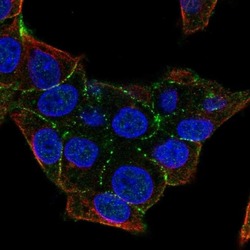
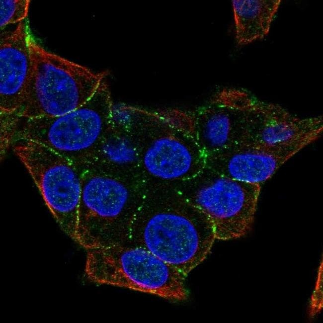
- Immunofluorescent staining of PKP4 in human cell line Hep G2 shows localization to cell junctions. Samples were probed using a PKP4 Polyclonal Antibody (Product # PA5-66855).
Supportive validation
- Submitted by
- Invitrogen Antibodies (provider)
- Main image
- Experimental details
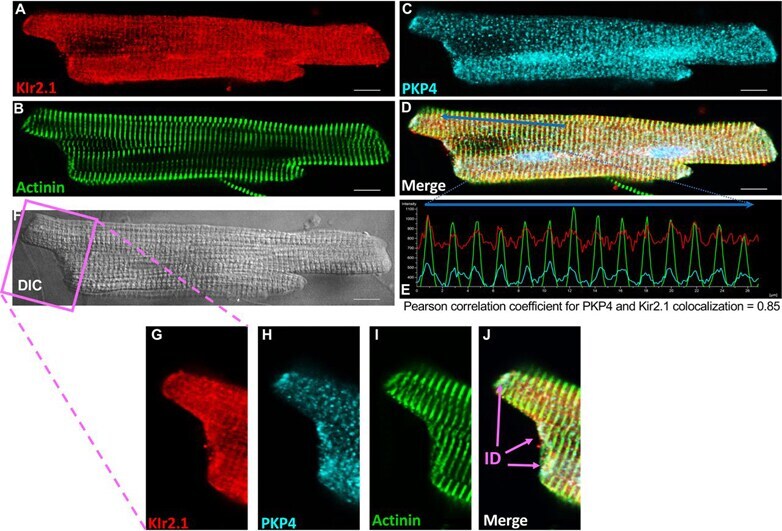
- Fig. 4 Kir2.1 and PKP4 co-localize in adult ventricular myocytes. Immunofluorescence (IF) staining analyses of the subcellular localization of Kir2.1 ( A , red ), Actinin ( B , green ) and PKP4 ( C , light blue ) in a freshly isolated rat adult ventricular myocytes. ( D ) Merge image. ( E ) Pixel intensity profile of PKP4, Kir2.1 and actinin along a line in the merge image, i.e. blue arrow shown in ( D ) showing the striated co-localization of the three proteins at the z-disks near the cardiac sarcomeres. ( F ) Differential interference contrast (DIC) image of the myocyte. ( G - J ) Zoomed in images of the intercalated disks. Scale bars: 10 mu m . ID: intercalated disk.