Explore
 Validate
 Learn
Learn Western blot
Antibody data
- Antibody Data
- Antigen structure
- References [0]
- Comments [0]
- Validations
- Western blot [1]
Submit
Validation data
Reference
Comment
Report error
- Product number
- A00422 - Provider product page

- Provider
- Boster Biological Technology
- Product name
- Anti-Fibroblast Activation Protein FAP Antibody
- Antibody type
- Polyclonal
- Description
- Polyclonal antibody for FAP detection. Host: Rabbit.Size: 100μL. Tested applications: IHC. Reactive species: Mouse. FAP information: Molecular Weight: 87713 MW; Subcellular Localization: Prolyl endopeptidase FAP: Cell surface . Cell membrane ; Single- pass type II membrane protein . Cell projection, lamellipodium membrane ; Single-pass type II membrane protein . Cell projection, invadopodium membrane ; Single-pass type II membrane protein . Cell projection, ruffle membrane ; Single-pass type II membrane protein . Membrane ; Single-pass type II membrane protein . Localized on cell surface with lamellipodia and invadopodia membranes and on shed vesicles. Colocalized with DPP4 at invadopodia and lamellipodia membranes of migratory activated endothelial cells in collagenous matrix. Colocalized with DPP4 on endothelial cells of capillary-like microvessels but not large vessels within invasive breast ductal carcinoma. Anchored and enriched preferentially by integrin alpha-3/beta-1 at invadopodia, plasma membrane protrusions that correspond to sites of cell invasion, in a collagen-dependent manner. Localized at plasma and ruffle membranes in a collagen-independent manner. Colocalized with PLAUR preferentially at the cell surface of invadopodia membranes in a cytoskeleton-, integrin- and vitronectin-dependent manner. Concentrated at invadopodia membranes, specialized protrusions of the ventral plasma membrane in a fibrobectin-dependent manner. Colocalizes with extracellular components (ECM), such as collagen fibers and fibronectin; Tissue Specificity: Expressed in adipose tissue. Expressed in the dermal fibroblasts in the fetal skin. Expressed in the granulation tissue of healing wounds and on reactive stromal fibroblast in epithelial cancers. Expressed in activated fibroblast-like synoviocytes from inflamed synovial tissues. Expressed in activated hepatic stellate cells (HSC) and myofibroblasts from cirrhotic liver, but not detected in normal liver. Expressed in glioma cells (at protein level). Expressed in glioblastomas and glioma cells. Isoform 1 and isoform 2 are expressed in melanoma, carcinoma and fibroblast cell lines.
- Reactivity
- Mouse
- Host
- Rabbit
- Vial size
- 100μL
- Concentration
- 1.1 mg/mL by UV absorbance at 280 nm
No comments: Submit comment
Supportive validation
- Submitted by
- Boster Biological Technology (provider)
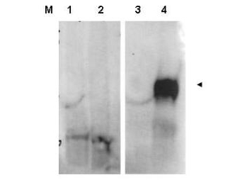
- Main image

- Experimental details
- Western blot analysis of FAP expression in whole cell lysates from FAP expressing HEK cells (lane 4) but not control HEK cells (lane 3). Specific band staining is blocked when the primary antibody is pre-incubated with immunizing peptide (lanes 1 and 2 respectively). FAP at 90KD was detected using rabbit anti-Fibroblast Activation Protein Antigen Affinity purified polyclonal antibody (Catalog # A00422) at 1:1000. Personal communication, S.Kim, NCI, Bethesda, MD.
- Additional image