 Explore
 Validate
 Learn
Learn Western blot
 Immunocytochemistry
Antibody data
- Antibody Data
- Antigen structure
- References [1]
- Comments [0]
- Validations
- Immunocytochemistry [8]
- Immunohistochemistry [1]
- Other assay [1]
Submit
Validation data
Reference
Comment
Report error
- Product number
- PA5-30428 - Provider product page

- Provider
- Invitrogen Antibodies
- Product name
- HSPE1 Polyclonal Antibody
- Antibody type
- Polyclonal
- Antigen
- Recombinant full-length protein
- Description
- Recommended positive controls: mouse brain. Predicted reactivity: Mouse (96%), Rat (98%), Zebrafish (82%), Japanese Medaka (86%), Xenopus laevis (88%), Pig (100%), Chicken (87%), Rhesus Monkey (99%), Bovine (100%). Store product as a concentrated solution. Centrifuge briefly prior to opening the vial.
- Reactivity
- Human, Mouse
- Host
- Rabbit
- Isotype
- IgG
- Vial size
- 100 μL
- Concentration
- 1 mg/mL
- Storage
- Store at 4°C short term. For long term storage, store at -20°C, avoiding freeze/thaw cycles.
Submitted references Oocytes maintain ROS-free mitochondrial metabolism by suppressing complex I.
Rodríguez-Nuevo A, Torres-Sanchez A, Duran JM, De Guirior C, Martínez-Zamora MA, Böke E
Nature 2022 Jul;607(7920):756-761
Nature 2022 Jul;607(7920):756-761
No comments: Submit comment
Supportive validation
- Submitted by
- Invitrogen Antibodies (provider)
- Main image

- Experimental details
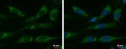
- Immunofluorescent analysis of HSPE1/Cpn10 showing staining in the mitochondria of SKNSH cells. SKNSH cells were fixed in 2% paraformaldehyde/culture medium at 37ºC for 15 min and stained using a HSPE1/Cpn10 polyclonal antibody (Product # PA5-30428) diluted at 1:500. Blue: Hoechst 33342 staining.
- Submitted by
- Invitrogen Antibodies (provider)
- Main image
- Experimental details
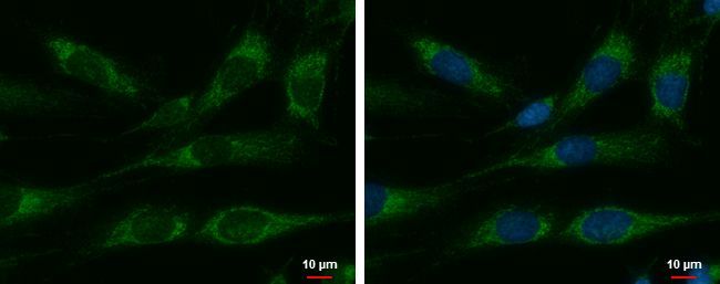
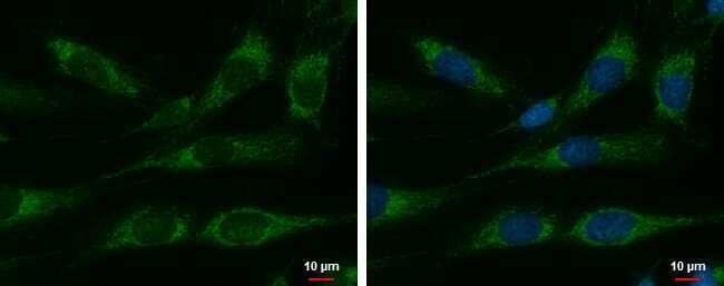
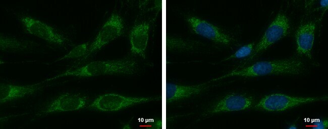
- HSPE1 Polyclonal Antibody detects Cpn10 protein at mitochondria by immunofluorescent analysis. Sample: SKNSH cells were fixed in 2% paraformaldehyde/culture medium at 37ºC for 15 min. Green: Cpn10 protein stained by HSPE1 Polyclonal Antibody (Product # PA5-30428) diluted at 1:500. Blue: Hoechst 33342 staining.
- Submitted by
- Invitrogen Antibodies (provider)
- Main image

- Experimental details
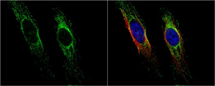
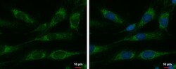
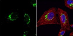
- Immunocytochemistry-Immunofluorescence analysis of HSPE1 was performed in HeLa cells fixed in 4% paraformaldehyde at RT for 15 min. Green: HSPE1 Polyclonal Antibody (Product # PA5-30428) diluted at 1:100. Red: alpha Tubulin, a cytoskeleton marker. Blue: Hoechst 33342 staining.
- Submitted by
- Invitrogen Antibodies (provider)
- Main image

- Experimental details
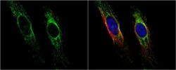
- Immunocytochemistry-Immunofluorescence analysis of HSPE1 was performed in U2OS cells fixed in 4% paraformaldehyde at RT for 15 min. Green: HSPE1 Polyclonal Antibody (Product # PA5-30428) diluted at 1:200. Red: alpha Tubulin, a cytoskeleton marker. Blue: Hoechst 33342 staining.

- Submitted by
- Invitrogen Antibodies (provider)
- Main image

- Experimental details
- HSPE1 Polyclonal Antibody detects Cpn10 protein at mitochondria by immunofluorescent analysis. Sample: SKNSH cells were fixed in 2% paraformaldehyde/culture medium at 37ºC for 15 min. Green: Cpn10 protein stained by HSPE1 Polyclonal Antibody (Product # PA5-30428) diluted at 1:500. Blue: Hoechst 33342 staining.
- Submitted by
- Invitrogen Antibodies (provider)
- Main image
- Experimental details
- HSPE1 Polyclonal Antibody detects Cpn10 protein at mitochondria by immunofluorescent analysis. Sample: SKNSH cells were fixed in 2% paraformaldehyde/culture medium at 37ºC for 15 min. Green: Cpn10 protein stained by HSPE1 Polyclonal Antibody (Product # PA5-30428) diluted at 1:500. Blue: Hoechst 33342 staining.

- Submitted by
- Invitrogen Antibodies (provider)
- Main image
- Experimental details
- Immunocytochemistry-Immunofluorescence analysis of HSPE1 was performed in HeLa cells fixed in 4% paraformaldehyde at RT for 15 min. Green: HSPE1 Polyclonal Antibody (Product # PA5-30428) diluted at 1:100. Red: alpha Tubulin, a cytoskeleton marker. Blue: Hoechst 33342 staining.

- Submitted by
- Invitrogen Antibodies (provider)
- Main image
- Experimental details
- Immunocytochemistry-Immunofluorescence analysis of HSPE1 was performed in U2OS cells fixed in 4% paraformaldehyde at RT for 15 min. Green: HSPE1 Polyclonal Antibody (Product # PA5-30428) diluted at 1:200. Red: alpha Tubulin, a cytoskeleton marker. Blue: Hoechst 33342 staining.

Supportive validation
- Submitted by
- Invitrogen Antibodies (provider)
- Main image
- Experimental details
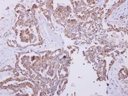
- HSPE1 Polyclonal Antibody detects Cpn10 protein at cytoplasm on human lung carcinoma by immunohistochemical analysis. Sample: Paraffin-embedded lung carcinoma. HSPE1 Polyclonal Antibody (Product # PA5-30428) dilution: 1:250. Antigen Retrieval: EDTA based buffer, pH 8.0, 15 min.

Supportive validation
- Submitted by
- Invitrogen Antibodies (provider)
- Main image
- Experimental details
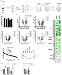
- Extended Data Fig. 3 OXPHOS machinery is reduced in Xenopus early oocytes. a , A schematic representation of isobaric-tag-based quantification of the mitochondrial proteomes of early (stage I), late (stage VI) oocytes, and muscle. The image was created with BioRender.com. b , Percentage of proteins identified in the isobaric-tag-based quantification compared to all known subunits of OXPHOS machinery belonging to complexes I to V. c , A volcano plot showing P values versus fold changes of mitochondrial proteins between early (stage I) and late (stage VI) oocytes. Proteins significantly changing ( q value 1.5-fold change) are depicted in black. Subunits of mitochondrial import machinery (TIMs and TOMs) are highlighted in light blue. n=3 biological replicates. d , A volcano plot showing P values versus fold changes of mitochondrial proteins between early (stage I) oocytes and muscle. Subunits of OXPHOS and mitochondrial import machinery are highlighted in indicated colours. Other proteins significantly changing ( q value 1.5-fold change) are depicted in black. n=3 biological replicates. e , Heatmap of fold changes (early- vs late-stage oocytes) of all quantified subunits of the OXPHOS and mitochondrial import machinery. # marks core subunits of complex I. f , g , h , Volcano plots displaying P values versus fold changes of mitochondrial proteins between early (stage I) oocytes and heart ( f ), liver ( g ) and white adipose tissue (WAT) ( h ). Subunits of OXPHOS a
