Explore
 Validate
 Learn
Learn Western blot
 Immunohistochemistry
Antibody data
- Antibody Data
- Antigen structure
- References [1]
- Comments [0]
- Validations
- Immunohistochemistry [2]
Submit
Validation data
Reference
Comment
Report error
- Product number
- PA5-22340 - Provider product page

- Provider
- Invitrogen Antibodies
- Product name
- GNAT2 Polyclonal Antibody
- Antibody type
- Polyclonal
- Antigen
- Recombinant full-length protein
- Description
- Predicted reactivity: Mouse (99%), Rat (99%), Zebrafish (87%), Xenopus laevis (91%), Chicken (94%), Bovine (99%). Store product as a concentrated solution. Centrifuge briefly prior to opening the vial.
- Reactivity
- Human, Mouse
- Host
- Rabbit
- Isotype
- IgG
- Vial size
- 100 μL
- Concentration
- 1.26 mg/mL
- Storage
- Store at 4°C short term. For long term storage, store at -20°C, avoiding freeze/thaw cycles.
Submitted references BBSome Component BBS5 Is Required for Cone Photoreceptor Protein Trafficking and Outer Segment Maintenance.
Bales KL, Bentley MR, Croyle MJ, Kesterson RA, Yoder BK, Gross AK
Investigative ophthalmology & visual science 2020 Aug 3;61(10):17
Investigative ophthalmology & visual science 2020 Aug 3;61(10):17
No comments: Submit comment
Supportive validation
- Submitted by
- Invitrogen Antibodies (provider)
- Main image
- Experimental details
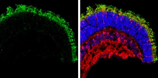
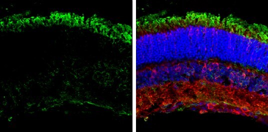
- Immunohistochemistry (Frozen) analysis of GNAT2 was performed in frozen sectioned adult mouse retina tissue using GNAT2 Polyclonal Antibody (Product # PA5-22340) at a dilution of 1:250 (Green). Red: beta Tubulin 3/ TUJ1, stained by beta Tubulin 3/ TUJ1 antibody diluted at 1:250. Blue: Fluoroshield with DAPI.
- Submitted by
- Invitrogen Antibodies (provider)
- Main image
- Experimental details
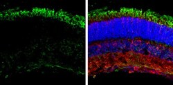
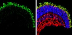
- Immunohistochemistry (Frozen) analysis of GNAT2 was performed in frozen sectioned adult mouse retina tissue using GNAT2 Polyclonal Antibody (Product # PA5-22340) at a dilution of 1:250 (Green). Red: beta Tubulin 3/ TUJ1, stained by beta Tubulin 3/ TUJ1 antibody diluted at 1:250. Blue: Fluoroshield with DAPI.