 Explore
 Validate
 Learn
Learn Western blot
 Immunohistochemistry
Antibody data
- Antibody Data
- Antigen structure
- References [2]
- Comments [0]
- Validations
- Immunohistochemistry [1]
- Flow cytometry [3]
- Other assay [2]
Submit
Validation data
Reference
Comment
Report error
- Product number
- PA5-26127 - Provider product page

- Provider
- Invitrogen Antibodies
- Product name
- AMPD2 Polyclonal Antibody
- Antibody type
- Polyclonal
- Antigen
- Synthetic peptide
- Reactivity
- Human
- Host
- Rabbit
- Isotype
- IgG
- Vial size
- 400 μL
- Concentration
- 2.0 mg/mL
- Storage
- Store at 4°C short term. For long term storage, store at -20°C, avoiding freeze/thaw cycles.
Submitted references The Anti-Glucocorticoid Receptor Antibody Clone 5E4: Raising Awareness of Unspecific Antibody Binding.
Surface AMP deaminase 2 as a novel regulator modifying extracellular adenine nucleotide metabolism.
Ehlers L, Kirchner M, Mertins P, Strehl C, Buttgereit F, Gaber T
International journal of molecular sciences 2022 May 2;23(9)
International journal of molecular sciences 2022 May 2;23(9)
Surface AMP deaminase 2 as a novel regulator modifying extracellular adenine nucleotide metabolism.
Ehlers L, Kuppe A, Damerau A, Wilantri S, Kirchner M, Mertins P, Strehl C, Buttgereit F, Gaber T
FASEB journal : official publication of the Federation of American Societies for Experimental Biology 2021 Jul;35(7):e21684
FASEB journal : official publication of the Federation of American Societies for Experimental Biology 2021 Jul;35(7):e21684
No comments: Submit comment
Supportive validation
- Submitted by
- Invitrogen Antibodies (provider)
- Main image
- Experimental details
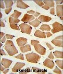
- Immunohistochemistry analysis of AMPD2 in formalin-fixed and paraffin-embedded human skeletal muscle. Samples were incubated with AMPD2 polyclonal antibody (Product # PA5-26127) which was peroxidase-conjugated to the secondary antibody, followed by DAB staining. This data demonstrates the use of this antibody for immunohistochemistry; clinical relevance has not been evaluated.

Supportive validation
- Submitted by
- Invitrogen Antibodies (provider)
- Main image

- Experimental details
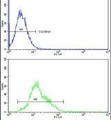
- Flow cytometry analysis of K562 cells using an AMPD2 polyclonal antibody (Product # PA5-26127) (bottom) compared to a negative control cell (top) at a dilution of 1:10-50, followed by a FITC-conjugated goat anti-rabbit antibody
- Submitted by
- Invitrogen Antibodies (provider)
- Main image
- Experimental details
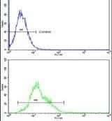
- Flow cytometry analysis of K562 cells using an AMPD2 polyclonal antibody (Product # PA5-26127) (bottom) compared to a negative control cell (top) at a dilution of 1:10-50, followed by a FITC-conjugated goat anti-rabbit antibody
- Submitted by
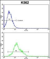
- Invitrogen Antibodies (provider)
- Main image

- Experimental details
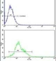
- Flow cytometry of AMPD2 in K562 cells (bottom histogram). Samples were incubated with AMPD2 polyclonal antibody (Product # PA5-26127) followed by FITC-conjugated goat-anti-rabbit secondary antibody. Negative control cell (top histogram).
Supportive validation
- Submitted by
- Invitrogen Antibodies (provider)
- Main image

- Experimental details
- Verification of anti-GR (5E4) antibody specificity. ( A ) Western blot analysis of GR pulled down from HEK293 membrane fractions by immunoprecipitation using the anti-GR (5E4) antibody (IP 5E4). An amount of 20 uL of membrane fraction protein (lysate) were analyzed in parallel. Protein detection was achieved by adding the anti-GR (5E4) antibody followed by an HRP-conjugated anti-mouse IgG antibody as a secondary reagent. ( B ) Immunoprecipitation from HEK293 membrane fractions was performed using the anti-GR (5E4) antibody (IP 5E4) and mouse IgG1 as a corresponding isotype control (IP IgG1). For mass spectrometric analysis the protein content was visualized by Pierce Coomassie Brilliant Blue G-250 Dye after SDS-PAGE, and the indicated area of interest was extracted for analysis. ( C ) Mass spectrometric analyses of pull-down samples obtained by immunoprecipitation from HEK293, Jurkat, and THP-1 whole cell lysates using the anti-GR (5E4) antibody (AB) and mouse IgG1 as corresponding isotype control (Control). Differential protein abundance compared to isotype control was determined using a two-sample Student's t test and black circles represent significance with an FDR cut-off of 5%. ( D ) Western blot analysis of GR pulled down from HEK293 whole cell lysates by immunoprecipitation using anti-GR (5E4) antibody Lot #1 (provided by Timea Berki []) and Lot #2 (Bio-Rad, Cat# MCA2469, RRID:AB_10844347). The protein content was visualized by incubation with anti-GR (5E4) antibody fo

- Submitted by
- Invitrogen Antibodies (provider)
- Main image
- Experimental details
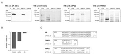
- Re-evaluation of anti-GR (5E4) antibody specificity. ( A ) Comparison of target proteins by western blot analysis. Pull-down samples from HEK293 whole cell lysates were obtained by immunoprecipitation using the following antibodies: anti-GR (5E4), anti-GR (G-5), anti-AMPD2 (QQ13), and anti-TRIM28 (Cat# PA5-27648). The protein content was visualized by incubation with primary antibodies directed against GR (5E4, biotinylated), GR (G-5, biotinylated), AMPD2 (PA5-26127, biotinylated), and TRIM28 (Cat# PA5-27648) as indicated, followed by HRP-conjugated streptavidin and anti-rabbit IgG antibody as secondary reagents. ( B ) Mass spectrometric analysis of pull-down samples obtained by IP from HEK293 whole cell lysates using the anti-GR antibody, 5E4, with and without prior two-hour incubation with APTEK-26 peptide. Bar graphs show fold change of peptide incubation to without peptide incubation. ( C ) Amino acid sequences of the newly identified anti-GR (5E4) target proteins, AMPD2 (UniProt ID: Q01433) and TRIM28 (UniProt ID: Q13263), were blasted against the APTEK-26 peptide using NCBI Protein BLAST ( https://blast.ncbi.nlm.nih.gov/Blast.cgi , accessed on 14 November 2020) [].
