Explore
 Validate
 Learn
LearnASC-002-25UL
antibody from Invitrogen Antibodies
Targeting: SCN2A
HBSCI, HBSCII, Nav1.2, SCN2A1, SCN2A2
 Western blot
 Immunoprecipitation
Antibody data
- Antibody Data
- Antigen structure
- References [0]
- Comments [0]
- Validations
- Western blot [2]
- Immunohistochemistry [1]
- Other assay [1]
Submit
Validation data
Reference
Comment
Report error
- Product number
- ASC-002-25UL - Provider product page

- Provider
- Invitrogen Antibodies
- Product name
- SCN2A (NaV1.2) Polyclonal Antibody
- Antibody type
- Polyclonal
- Antigen
- Other
- Reactivity
- Human, Mouse, Rat
- Host
- Rabbit
- Isotype
- IgG
- Vial size
- 25 µL
- Concentration
- 0.8 mg/mL
- Storage
- -20° C, Avoid Freeze/Thaw Cycles
No comments: Submit comment
Supportive validation
- Submitted by
- Invitrogen Antibodies (provider)
- Main image
- Experimental details
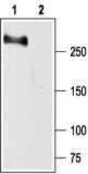
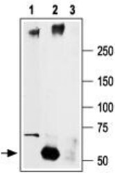
- Western blot analysisof rat brain membranes: - 1. Anti-SCN2A (NaV1.2) Antibody (#ASC-002), (1:200). 2. Anti-SCN2A (NaV1.2) Antibody , preincubated with SCN2A/Nav1.2 Blocking Peptide (#BLP-SC002).

- Submitted by
- Invitrogen Antibodies (provider)
- Main image
- Experimental details
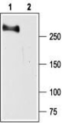
- Western blot analysisof rat brain membranes: - 1. Anti-SCN2A (NaV1.2) Antibody (#ASC-002), (1:200). 2. Anti-SCN2A (NaV1.2) Antibody , preincubated with SCN2A/Nav1.2 Blocking Peptide (#BLP-SC002).

Supportive validation
- Submitted by
- Invitrogen Antibodies (provider)
- Main image
- Experimental details
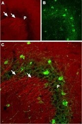
- Expression of NaV1.2 in mouse hippocampus - Immunohistochemical staining of mouse hippocampus using Anti-SCN2A (NaV1.2) Antibody (#ASC-002 ). A. NaV1.2 (red) is present in dendrites of pyramidal neurons in the CA3 region. B. Staining ofinterneurons in the pyramidal layer with mouse Anti-Parvalbumin (green) demonstrates the restriction of NaV1.2 to dendrites (arrows) extending from the pyramidal layer (P). C. Confocal merge of panels A and B.

Supportive validation
- Submitted by
- Invitrogen Antibodies (provider)
- Main image
- Experimental details
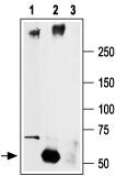
- Immunoprecipitation of rat brain lysate: - 1. Rat brain lysates2. Lysates immunoprecipitated with Anti-SCN2A (NaV1.2) Antibody (#ASC-002), (6 µg).3. Lysates immunoprecipitated with pre-immune rabbit serum. The lower arrow indicates the IgG heavy chain. Western blot analysiswas performed with Anti-SCN2A (NaV1.2) Antibody .