Explore
 Validate
 Learn
Learn Western blot
 Immunocytochemistry
Antibody data
- Antibody Data
- Antigen structure
- References [2]
- Comments [0]
- Validations
- Immunocytochemistry [8]
- Flow cytometry [1]
- Other assay [1]
Submit
Validation data
Reference
Comment
Report error
- Product number
- PA1-007 - Provider product page

- Provider
- Invitrogen Antibodies
- Product name
- ERp72 Polyclonal Antibody
- Antibody type
- Polyclonal
- Antigen
- Synthetic peptide
- Description
- PA1-007 detects ERp72 protein in human and mouse samples. PA1-007 has successfully been used in ICC, IF and Western blot procedures. By Western blot, this antibody detects a 72 kDa protein representing ERp72 in human fibrosarcoma HT 1080 cell lysate. The PA1-007 immunogen is a synthetic peptide corresponding to residues K(629) F I E E H A T K L S R T K E E L(645) of humna ERp72. This peptide (Cat. # PEP-223) is available for use in neutralization and control experiments.
- Reactivity
- Human, Mouse
- Host
- Rabbit
- Isotype
- IgG
- Vial size
- 100 μg
- Concentration
- 1 mg/mL
- Storage
- -20°C, Avoid Freeze/Thaw Cycles
Submitted references Protein Disulfide Isomerase A4 Is Involved in Genome Uncoating during Human Astrovirus Cell Entry.
Axons provide the secretory machinery for trafficking of voltage-gated sodium channels in peripheral nerve.
Aguilar-Hernández N, Meyer L, López S, DuBois RM, Arias CF
Viruses 2020 Dec 31;13(1)
Viruses 2020 Dec 31;13(1)
Axons provide the secretory machinery for trafficking of voltage-gated sodium channels in peripheral nerve.
González C, Cánovas J, Fresno J, Couve E, Court FA, Couve A
Proceedings of the National Academy of Sciences of the United States of America 2016 Feb 16;113(7):1823-8
Proceedings of the National Academy of Sciences of the United States of America 2016 Feb 16;113(7):1823-8
No comments: Submit comment
Supportive validation
- Submitted by
- Invitrogen Antibodies (provider)
- Main image
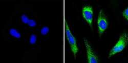
- Experimental details
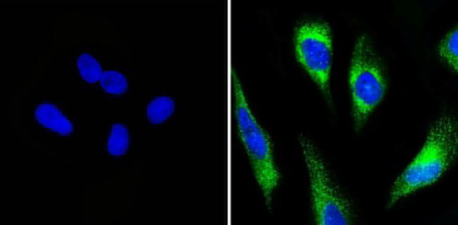
- Immunofluorescent analysis of ERp72 (green) showing ER staining in Hela cells (right) compared to a negative control without primary antibody (left). Cells were permeabilized with 0.1% Triton X-100 in TBS for 5-10 minutes and blocked with 3% BSA-PBS for 30 minutes at room temperature. Cells were probed with a ERp72 polyclonal antibody (Product # PA1-007) in 3% BSA-PBS at a dilution of 1:100 and incubated overnight at 4ºC in a humidified chamber. Cells were washed with PBST and incubated with a DyLight-488 conjugated secondary antibody in PBS at room temperature in the dark. Nuclei were stained with DAPI (blue) and images were taken at a magnification of 60x.
- Submitted by
- Invitrogen Antibodies (provider)
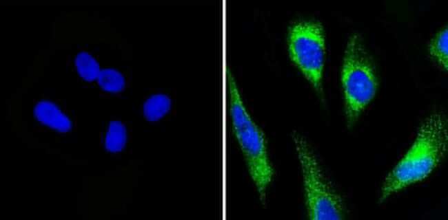
- Main image

- Experimental details
- Immunofluorescent analysis of ERp72 (green) showing ER staining in MCF-7 cells (right) compared to a negative control without primary antibody (left). Cells were permeabilized with 0.1% Triton X-100 in TBS for 5-10 minutes and blocked with 3% BSA-PBS for 30 minutes at room temperature. Cells were probed with a ERp72 polyclonal antibody (Product # PA1-007) in 3% BSA-PBS at a dilution of 1:100 and incubated overnight at 4ºC in a humidified chamber. Cells were washed with PBST and incubated with a DyLight-488 conjugated secondary antibody in PBS at room temperature in the dark. Nuclei were stained with DAPI (blue) and images were taken at a magnification of 60x.
- Submitted by
- Invitrogen Antibodies (provider)
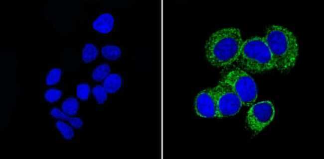
- Main image

- Experimental details
- Immunofluorescent analysis of ERp72 (green) showing ER staining in NIH-3T3 cells (right) compared to a negative control without primary antibody (left). Cells were permeabilized with 0.1% Triton X-100 in TBS for 5-10 minutes and blocked with 3% BSA-PBS for 30 minutes at room temperature. Cells were probed with a ERp72 polyclonal antibody (Product # PA1-007) in 3% BSA-PBS at a dilution of 1:100 and incubated overnight at 4ºC in a humidified chamber. Cells were washed with PBST and incubated with a DyLight-488 conjugated secondary antibody in PBS at room temperature in the dark. Actin was stained with a Dylight 554 phalloidin (red) and nuclei were stained with DAPI (blue). Images were taken at a magnification of 60x.

- Submitted by
- Invitrogen Antibodies (provider)
- Main image

- Experimental details
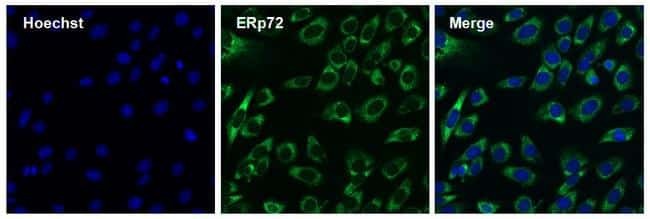
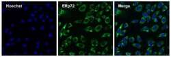
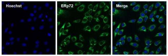
- Immunofluorescent analysis of ERp72 (green) in 3T3 cells. The cells were permeabilized with 0.1% Triton X-100 in TBS for 15 minutes, and blocked with 3% Blocker BSA in PBS (Product # 37525) for 15 minutes at room temperature. Cells were stained with a ERp72 rabbit polyclonal antibody (Product # PA1-007), at a concentration of 10 µg/mL in blocking buffer for at least 1 hour at room temperature, and then incubated with a Goat anti-rabbit IgG Superclonal secondary antibody, Alexa Fluor 488 conjugate (Product # A27034) at a dilution of 1:1000 for 30 minutes at room temperature (green). Nuclei (blue) were stained with Hoechst 33342 dye (Product # 62249). Images were taken on a Thermo Scientific ToxInsight Instrument at 20X magnification.
- Submitted by
- Invitrogen Antibodies (provider)
- Main image

- Experimental details
- Immunofluorescent analysis of ERp72 (green) showing ER staining in Hela cells (right) compared to a negative control without primary antibody (left). Cells were permeabilized with 0.1% Triton X-100 in TBS for 5-10 minutes and blocked with 3% BSA-PBS for 30 minutes at room temperature. Cells were probed with a ERp72 polyclonal antibody (Product # PA1-007) in 3% BSA-PBS at a dilution of 1:100 and incubated overnight at 4ºC in a humidified chamber. Cells were washed with PBST and incubated with a DyLight-488 conjugated secondary antibody in PBS at room temperature in the dark. Nuclei were stained with DAPI (blue) and images were taken at a magnification of 60x.
- Submitted by
- Invitrogen Antibodies (provider)
- Main image
- Experimental details
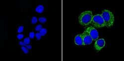
- Immunofluorescent analysis of ERp72 (green) showing ER staining in MCF-7 cells (right) compared to a negative control without primary antibody (left). Cells were permeabilized with 0.1% Triton X-100 in TBS for 5-10 minutes and blocked with 3% BSA-PBS for 30 minutes at room temperature. Cells were probed with a ERp72 polyclonal antibody (Product # PA1-007) in 3% BSA-PBS at a dilution of 1:100 and incubated overnight at 4ºC in a humidified chamber. Cells were washed with PBST and incubated with a DyLight-488 conjugated secondary antibody in PBS at room temperature in the dark. Nuclei were stained with DAPI (blue) and images were taken at a magnification of 60x.

- Submitted by
- Invitrogen Antibodies (provider)
- Main image
- Experimental details
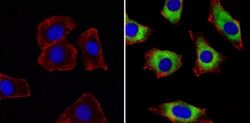
- Immunofluorescent analysis of ERp72 (green) showing ER staining in NIH-3T3 cells (right) compared to a negative control without primary antibody (left). Cells were permeabilized with 0.1% Triton X-100 in TBS for 5-10 minutes and blocked with 3% BSA-PBS for 30 minutes at room temperature. Cells were probed with a ERp72 polyclonal antibody (Product # PA1-007) in 3% BSA-PBS at a dilution of 1:100 and incubated overnight at 4ºC in a humidified chamber. Cells were washed with PBST and incubated with a DyLight-488 conjugated secondary antibody in PBS at room temperature in the dark. Actin was stained with a Dylight 554 phalloidin (red) and nuclei were stained with DAPI (blue). Images were taken at a magnification of 60x.

- Submitted by
- Invitrogen Antibodies (provider)
- Main image
- Experimental details
- Immunofluorescent analysis of ERp72 (green) in 3T3 cells. The cells were permeabilized with 0.1% Triton X-100 in TBS for 15 minutes, and blocked with 3% Blocker BSA in PBS (Product # 37525) for 15 minutes at room temperature. Cells were stained with a ERp72 rabbit polyclonal antibody (Product # PA1-007), at a concentration of 10 µg/mL in blocking buffer for at least 1 hour at room temperature, and then incubated with a Goat anti-rabbit IgG Superclonal secondary antibody, Alexa Fluor 488 conjugate (Product # A27034) at a dilution of 1:1000 for 30 minutes at room temperature (green). Nuclei (blue) were stained with Hoechst 33342 dye (Product # 62249). Images were taken on a Thermo Scientific ToxInsight Instrument at 20X magnification.
Supportive validation
- Submitted by
- Invitrogen Antibodies (provider)
- Main image
- Experimental details
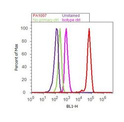
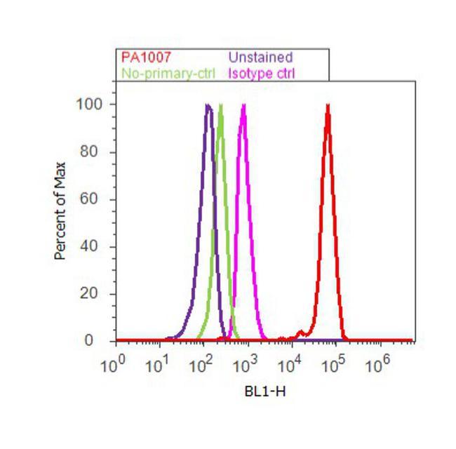
- Flow cytometry analysis of ER [p72] was done on Hep G2 cells. Cells were fixed with 70% ethanol for 10 minutes, permeabilized with 0.25% Triton™ X-100 for 20 minutes, and blocked with 5% BSA for 30 minutes at room temperature. Cells were labeled with ER [p72] Rabbit Polyclonal Antibody (PA1-007, red histogram) or with rabbit isotype control (pink histogram) at 3-5 ug/million cells in 2.5% BSA. After incubation at room temperature for 2 hours, the cells were labeled with Alexa Fluor® 488 Goat Anti-Rabbit Secondary Antibody (A11008) at a dilution of 1:400 for 30 minutes at room temperature. The representative 10, 000 cells were acquired and analyzed for each sample using an Attune® Acoustic Focusing Cytometer. The purple histogram represents unstained control cells and the green histogram represents no-primary-antibody control.
Supportive validation
- Submitted by
- Invitrogen Antibodies (provider)
- Main image
- Experimental details
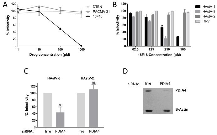
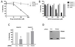
- Figure 4 PDIA4 is important for astrovirus infection. ( A ) Caco-2 cells were incubated with the indicated PDI inhibitors for 1 h at 37 degC, the cells were washed and infected with HAstV-8 and 16 h post-infection (hpi), and the viral infectivity was determined by an immunoperoxidase assay as described in Material and Methods. ( B ) Caco-2 cells were pre-treated for 1 h at 37 degC with the indicated concentrations of PDI inhibitor 16F16. Cells were washed and infected with either HAstV-1, -2, -8 or with rhesus rotavirus (RRV), and the viral infectivity was determined. Data in A and B are expressed as percentage of viral infectivity compared to control cells treated with DMSO, which was taken as 100%. ( C ) Caco-2 cells were transfected with an siRNA pool directed to PDIA4 or with an irrelevant (Irre) siRNA pool, and 72 h post-transfection (hpt) cells were infected with the indicated HAstV strain (MOI = 0.025). At 16 hpi, the cells were fixed, and the viral infectivity was determined as described previously. Virus infectivity is expressed as the percentage of infected cells obtained in the control-transfected cells (Irre), which was taken as 100%. The arithmetic mean +- SEM from three independent experiments performed in duplicate is shown. * p < 0.05. ( D ) A representative Western blot showing the effect of the siRNA-PDIA4 on the expression of PDIA4 as compared to cells transfected with an irrelevant siRNA. beta-Actin was used as a loading control.