Explore
 Validate
 Learn
Learn Immunocytochemistry
Antibody data
- Antibody Data
- Antigen structure
- References [2]
- Comments [0]
- Validations
- Immunocytochemistry [1]
- Immunohistochemistry [1]
Submit
Validation data
Reference
Comment
Report error
- Product number
- HPA051467 - Provider product page

- Provider
- Atlas Antibodies
- Proper citation
- Atlas Antibodies Cat#HPA051467, RRID:AB_2681495
- Product name
- Anti-LAMP3
- Antibody type
- Polyclonal
- Description
- Polyclonal Antibody against Human LAMP3, Gene description: lysosomal-associated membrane protein 3, Alternative Gene Names: CD208, DC-LAMP, DCLAMP, LAMP, TSC403, Validated applications: ICC, IHC, Uniprot ID: Q9UQV4, Storage: Store at +4°C for short term storage. Long time storage is recommended at -20°C.
- Reactivity
- Human
- Host
- Rabbit
- Conjugate
- Unconjugated
- Isotype
- IgG
- Vial size
- 100 µl
- Concentration
- 0.1 mg/ml
- Storage
- Store at +4°C for short term storage. Long time storage is recommended at -20°C.
- Handling
- The antibody solution should be gently mixed before use.
Submitted references High-Resolution Analysis of Mononuclear Phagocytes Reveals GPNMB as a Prognostic Marker in Human Colorectal Liver Metastasis
Pancreatic Cancer Chemotherapy Is Potentiated by Induction of Tertiary Lymphoid Structures in Mice
Cortese N, Carriero R, Barbagallo M, Putignano A, Costa G, Giavazzi F, Grizzi F, Pasqualini F, Peano C, Basso G, Marchini S, Colombo F, Soldani C, Franceschini B, Di Tommaso L, Terracciano L, Donadon M, Torzilli G, Kunderfranco P, Mantovani A, Marchesi F
Cancer Immunology Research 2023;11(4):405-420
Cancer Immunology Research 2023;11(4):405-420
Pancreatic Cancer Chemotherapy Is Potentiated by Induction of Tertiary Lymphoid Structures in Mice
Delvecchio F, Fincham R, Spear S, Clear A, Roy-Luzarraga M, Balkwill F, Gribben J, Bombardieri M, Hodivala-Dilke K, Capasso M, Kocher H
Cellular and Molecular Gastroenterology and Hepatology 2021;12(5):1543-1565
Cellular and Molecular Gastroenterology and Hepatology 2021;12(5):1543-1565
No comments: Submit comment
Supportive validation
- Submitted by
- Atlas Antibodies (provider)
- Main image
- Experimental details
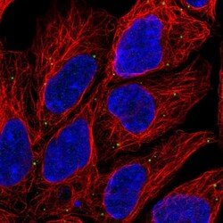
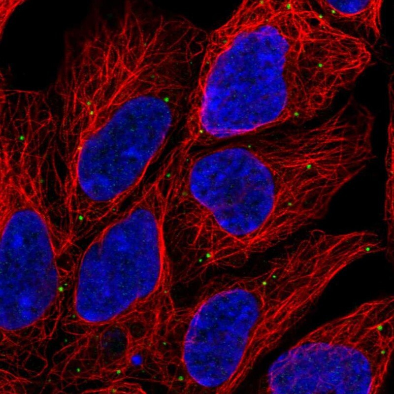
- Immunofluorescent staining of human cell line HEK 293 shows localization to vesicles.
- Sample type
- Human
Supportive validation
- Submitted by
- Atlas Antibodies (provider)
- Enhanced method
- Orthogonal validation
- Main image
- Experimental details
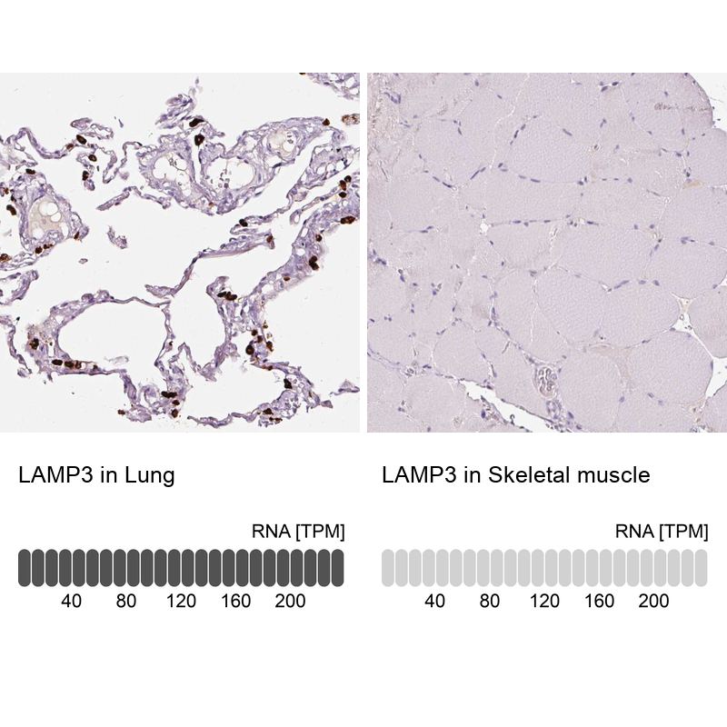
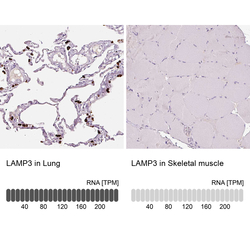
- Immunohistochemistry analysis in human lung and skeletal muscle tissues using HPA051467 antibody. Corresponding LAMP3 RNA-seq data are presented for the same tissues.
- Sample type
- Human
- Protocol
- Protocol