 Explore
 Validate
 Learn
Learn Western blot
 Immunocytochemistry
Antibody data
- Antibody Data
- Antigen structure
- References [8]
- Comments [0]
- Validations
- Immunocytochemistry [4]
- Other assay [4]
Submit
Validation data
Reference
Comment
Report error
- Product number
- MA3-413 - Provider product page

- Provider
- Invitrogen Antibodies
- Product name
- HIP Monoclonal Antibody (2G6)
- Antibody type
- Monoclonal
- Antigen
- Recombinant full-length protein
- Description
- MA3-413 detects heat shock cognate protein 70 kDa (HSC70)-interacting protein (Hip) from human, mouse, chicken and rabbit samples. MA3-413 has been successfully used in Western blot, immunofluorescence, and immunoprecipitation procedures. By Western blot, this antibody detects a 48 kDa protein representing Hip from rabbit reticulocyte lysate. The MA3-413 antigen is recombinant human Hip protein.
- Reactivity
- Human, Mouse, Chicken/Avian, Rabbit
- Host
- Mouse
- Isotype
- IgG
- Antibody clone number
- 2G6
- Vial size
- 50 µL
- Concentration
- Conc. Not Determined
- Storage
- -20° C, Avoid Freeze/Thaw Cycles
Submitted references Heat Treatment at an Early Age Has Effects on the Resistance to Chronic Heat Stress on Broilers.
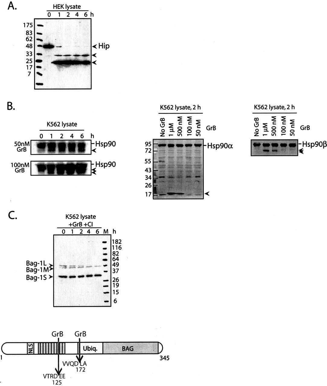
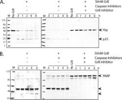
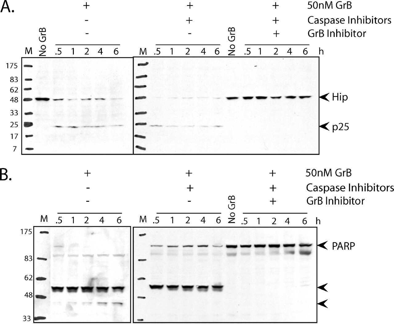
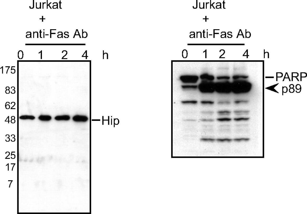
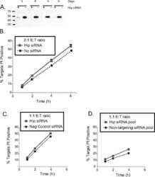
Hip is a pro-survival substrate of granzyme B.
hsp70 interacting protein Hip does not affect glucocorticoid receptor folding by the hsp90-based chaperone machinery except to oppose the effect of BAG-1.
Differential effects of the hsp70-binding protein BAG-1 on glucocorticoid receptor folding by the hsp90-based chaperone machinery.
Differential effects of the hsp70-binding protein BAG-1 on glucocorticoid receptor folding by the hsp90-based chaperone machinery.
Mutation of Hip's carboxy-terminal region inhibits a transitional stage of progesterone receptor assembly.
Molecular cloning of human p48, a transient component of progesterone receptor complexes and an Hsp70-binding protein.
Hip, a novel cochaperone involved in the eukaryotic Hsc70/Hsp40 reaction cycle.
Kang D, Park J, Shim K
Animals : an open access journal from MDPI 2019 Nov 23;9(12)
Animals : an open access journal from MDPI 2019 Nov 23;9(12)
Hip is a pro-survival substrate of granzyme B.
Hostetter DR, Loeb CR, Chu F, Craik CS
The Journal of biological chemistry 2007 Sep 21;282(38):27865-74
The Journal of biological chemistry 2007 Sep 21;282(38):27865-74
hsp70 interacting protein Hip does not affect glucocorticoid receptor folding by the hsp90-based chaperone machinery except to oppose the effect of BAG-1.
Kanelakis KC, Murphy PJ, Galigniana MD, Morishima Y, Takayama S, Reed JC, Toft DO, Pratt WB
Biochemistry 2000 Nov 21;39(46):14314-21
Biochemistry 2000 Nov 21;39(46):14314-21
Differential effects of the hsp70-binding protein BAG-1 on glucocorticoid receptor folding by the hsp90-based chaperone machinery.
Kanelakis KC, Morishima Y, Dittmar KD, Galigniana MD, Takayama S, Reed JC, Pratt WB
The Journal of biological chemistry 1999 Nov 26;274(48):34134-40
The Journal of biological chemistry 1999 Nov 26;274(48):34134-40
Mutation of Hip's carboxy-terminal region inhibits a transitional stage of progesterone receptor assembly.
Prapapanich V, Chen S, Smith DF
Molecular and cellular biology 1998 Feb;18(2):944-52
Molecular and cellular biology 1998 Feb;18(2):944-52
Molecular cloning of human p48, a transient component of progesterone receptor complexes and an Hsp70-binding protein.
Prapapanich V, Chen S, Nair SC, Rimerman RA, Smith DF
Molecular endocrinology (Baltimore, Md.) 1996 Apr;10(4):420-31
Molecular endocrinology (Baltimore, Md.) 1996 Apr;10(4):420-31
Hip, a novel cochaperone involved in the eukaryotic Hsc70/Hsp40 reaction cycle.
Höhfeld J, Minami Y, Hartl FU
Cell 1995 Nov 17;83(4):589-98
Cell 1995 Nov 17;83(4):589-98
No comments: Submit comment
Supportive validation
- Submitted by
- Invitrogen Antibodies (provider)
- Main image

- Experimental details
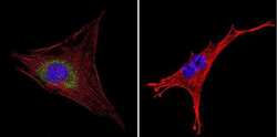
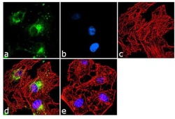
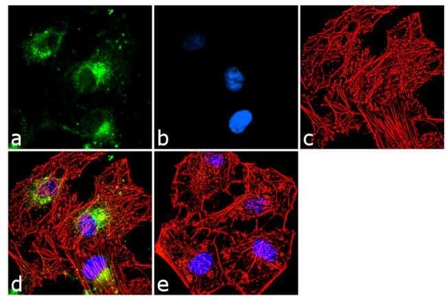
- Immunofluorescent analysis of HIP in A431 cells. Cells were grown on chamber slides and fixed with formaldehyde prior to staining. Cells were probed without (control) or with a HIP monoclonal antibody (Product # MA3-413) at a dilution of 1:100 overnight at 4 C, washed with PBS and incubated with a DyLight-488 conjugated secondary antibody (Product # 35503). HIP staining (green), F-Actin staining with Phalloidin (red) and nuclei with DAPI (blue) is shown. Images were taken at 60X magnification.
- Submitted by
- Invitrogen Antibodies (provider)
- Main image
- Experimental details
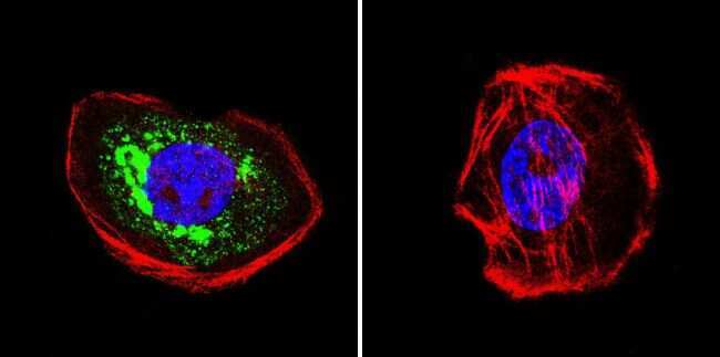
- Immunofluorescent analysis of HIP in HeLa cells. Cells were grown on chamber slides and fixed with formaldehyde prior to staining. Cells were probed without (control) or with a HIP monoclonal antibody (Product # MA3-413) at a dilution of 1:20 overnight at 4 C, washed with PBS and incubated with a DyLight-488 conjugated secondary antibody (Product # 35503). HIP staining (green), F-Actin staining with Phalloidin (red) and nuclei with DAPI (blue) is shown. Images were taken at 60X magnification.

- Submitted by
- Invitrogen Antibodies (provider)
- Main image
- Experimental details
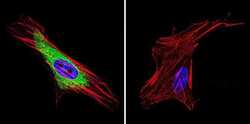
- Immunofluorescent analysis of HIP in NIH-3T3 cells. Cells were grown on chamber slides and fixed with formaldehyde prior to staining. Cells were probed without (control) or with a HIP monoclonal antibody (Product # MA3-413) at a dilution of 1:200 overnight at 4 C, washed with PBS and incubated with a DyLight-488 conjugated secondary antibody (Product # 35503). HIP staining (green), F-Actin staining with Phalloidin (red) and nuclei with DAPI (blue) is shown. Images were taken at 60X magnification.
- Submitted by
- Invitrogen Antibodies (provider)
- Main image
- Experimental details
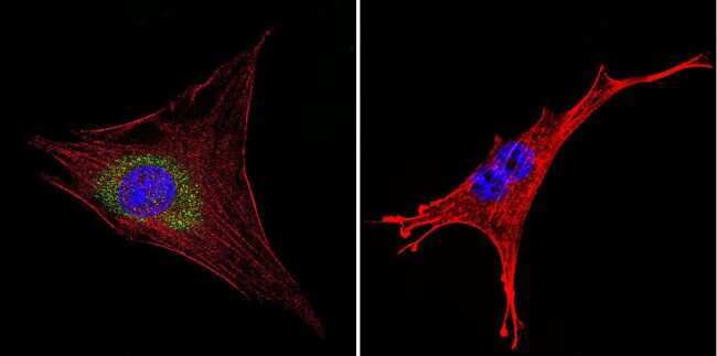
- Immunofluorescence analysis of HSC70 Interacting Protein (HIP) was done on 70% confluent log phase HeLa cells. The cells were fixed with 4% paraformaldehyde for 10 minutes, permeabilized with 0.1% Triton™ X-100 for 10 minutes, and blocked with 1% BSA for 1 hour at room temperature. The cells were labeled HSC70 Interacting Protein HIP (2G6) Mouse Monoclonal Antibody (Product # MA3-413) at 1:250 dilution in 0.1% BSA and incubated for 3 hours at room temperature and then labeled with Goat anti-Mouse IgG (H+L) Superclonal™ Secondary Antibody, Alexa Fluor® 488 conjugate (Product # A28175) at a dilution of 1:2000 for 45 minutes at room temperature (Panel a: green). Nuclei (Panel b: blue) were stained with SlowFade® Gold Antifade Mountant with DAPI (Product # S36938). F-actin (Panel c: red) was stained with Rhodamine Phalloidin (Product # R415, 1:300). Panel d is a merged image showing cytoplasmic localization. Panel e is a no primary antibody control. The images were captured at 60X magnification.
Supportive validation
- Submitted by
- Invitrogen Antibodies (provider)
- Main image

- Experimental details
- NULL
- Submitted by
- Invitrogen Antibodies (provider)
- Main image
- Experimental details
- NULL
- Submitted by
- Invitrogen Antibodies (provider)
- Main image

- Experimental details
- NULL
- Submitted by
- Invitrogen Antibodies (provider)
- Main image
- Experimental details
- NULL
