Explore
 Validate
 Learn
Learn Western blot
 Immunohistochemistry
Antibody data
- Antibody Data
- Antigen structure
- References [2]
- Comments [0]
- Validations
- Western blot [1]
- Immunocytochemistry [2]
Submit
Validation data
Reference
Comment
Report error
- Product number
- HPA035275 - Provider product page

- Provider
- Atlas Antibodies
- Proper citation
- Atlas Antibodies Cat#HPA035275, RRID:AB_10602600
- Product name
- Anti-GORASP2
- Antibody type
- Polyclonal
- Description
- Polyclonal Antibody against Human GORASP2, Gene description: golgi reassembly stacking protein 2, 55kDa, Alternative Gene Names: GOLPH6, GRASP55, GRS2, Validated applications: ICC, IHC, WB, Uniprot ID: Q9H8Y8, Storage: Store at +4°C for short term storage. Long time storage is recommended at -20°C.
- Reactivity
- Human, Mouse
- Host
- Rabbit
- Conjugate
- Unconjugated
- Isotype
- IgG
- Vial size
- 100 µl
- Concentration
- 0.1 mg/ml
- Storage
- Store at +4°C for short term storage. Long time storage is recommended at -20°C.
- Handling
- The antibody solution should be gently mixed before use.
Submitted references Muscle Proteomic Profile before and after Enzyme Replacement Therapy in Late-Onset Pompe Disease
Granulovacuolar degeneration bodies are neuron-selective lysosomal structures induced by intracellular tau pathology.
Moriggi M, Capitanio D, Torretta E, Barbacini P, Bragato C, Sartori P, Moggio M, Maggi L, Mora M, Gelfi C
International Journal of Molecular Sciences 2021;22(6):2850
International Journal of Molecular Sciences 2021;22(6):2850
Granulovacuolar degeneration bodies are neuron-selective lysosomal structures induced by intracellular tau pathology.
Wiersma VI, van Ziel AM, Vazquez-Sanchez S, Nölle A, Berenjeno-Correa E, Bonaterra-Pastra A, Clavaguera F, Tolnay M, Musters RJP, van Weering JRT, Verhage M, Hoozemans JJM, Scheper W
Acta neuropathologica 2019 Dec;138(6):943-970
Acta neuropathologica 2019 Dec;138(6):943-970
No comments: Submit comment
Enhanced validation
- Submitted by
- Atlas Antibodies (provider)
- Enhanced method
- Genetic validation
- Main image

- Experimental details
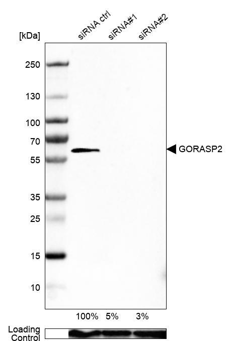
- Western blot analysis in U-251MG cells transfected with control siRNA, target specific siRNA probe #1 and #2, using Anti-GORASP2 antibody. Remaining relative intensity is presented. Loading control: Anti-GAPDH.
- Sample type
- Human
- Protocol
- Protocol
Enhanced validation
Supportive validation
- Submitted by
- 55af80e3e0991
- Enhanced method
- Genetic validation
- Main image
- Experimental details
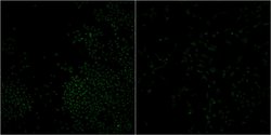
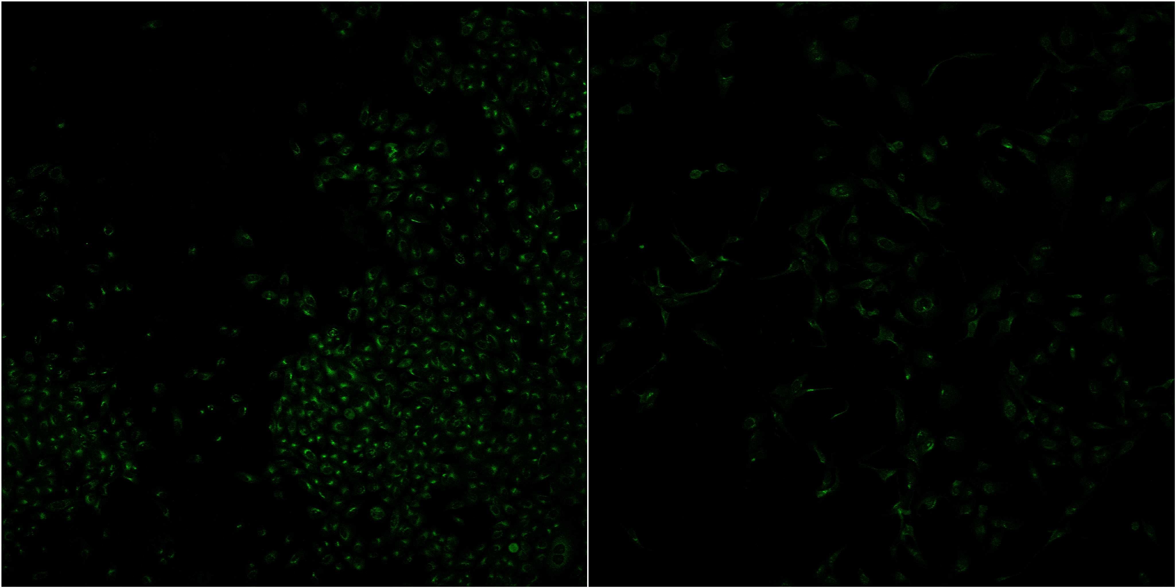
- Confocal images of immunofluorescently stained human U-2 OS cells.The protein GORASP2 is shown in green. The image to the left show cells transfected with control siRNA and the image to the right show cells where GORASP2 has been downregulated with specific siRNA.
- Sample type
- U-2 OS cells
- Primary Ab dilution
- 1:43
- Secondary Ab
- Secondary Ab
- Secondary Ab dilution
- 1:800
- Knockdown/Genetic Approaches Application
- Immunocytochemistry
Supportive validation
- Submitted by
- Atlas Antibodies (provider)
- Main image

- Experimental details
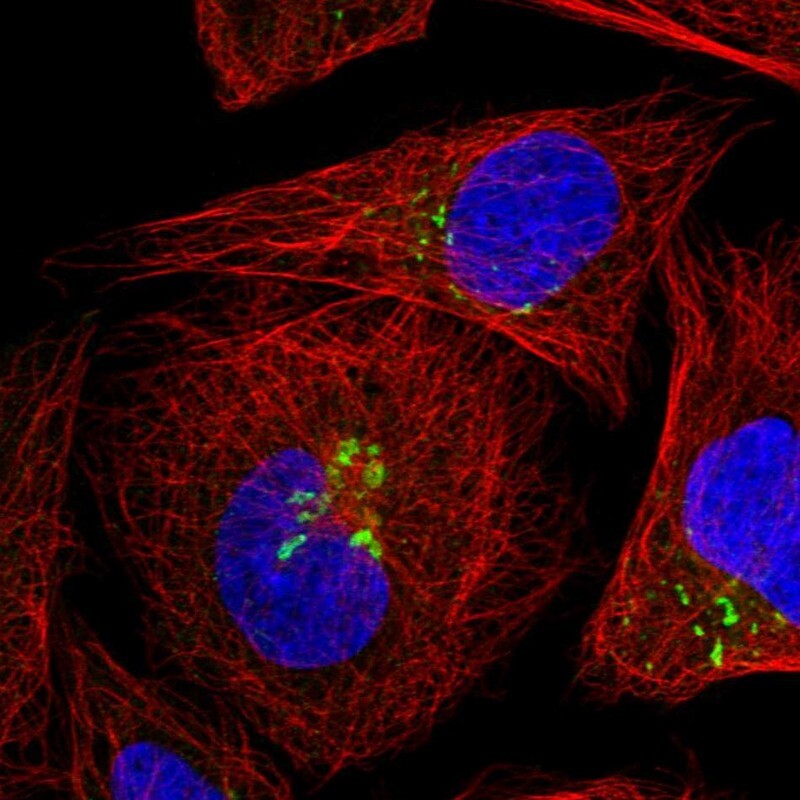
- Immunofluorescent staining of human cell line U-2 OS shows localization to the Golgi apparatus.
- Sample type
- Human