Explore
 Validate
 Learn
Learn Western blot
 Immunocytochemistry
Antibody data
- Antibody Data
- Antigen structure
- References [2]
- Comments [0]
- Validations
- Western blot [1]
- Immunocytochemistry [1]
- Immunohistochemistry [1]
Submit
Validation data
Reference
Comment
Report error
- Product number
- AMAb91016 - Provider product page

- Provider
- Atlas Antibodies
- Proper citation
- Atlas Antibodies Cat#AMAb91016, RRID:AB_2665764
- Product name
- Anti-GORASP2
- Antibody type
- Monoclonal
- Description
- Monoclonal Antibody against Human GORASP2, Clone ID: CL2610, Gene description: Golgi reassembly stacking protein 2, Alternative Gene Names: GOLPH6, GRASP55, GRS2, Validated applications: WB, IHC, ICC, Uniprot ID: Q9H8Y8, Storage: Store at +4°C for short term storage. Long time storage is recommended at -20°C.
- Reactivity
- Human
- Host
- Mouse
- Conjugate
- Unconjugated
- Isotype
- IgG
- Antibody clone number
- CL2610
- Vial size
- 100 µl
- Concentration
- 1.0 mg/ml
- Storage
- Store at +4°C for short term storage. Long time storage is recommended at -20°C.
- Handling
- The antibody solution should be gently mixed before use.
Submitted references Prenylcysteine oxidase 1, an emerging player in atherosclerosis
A Genome-Wide Knockout Screen in Human Macrophages Identified Host Factors Modulating Salmonella Infection
Banfi C, Baetta R, Barbieri S, Brioschi M, Guarino A, Ghilardi S, Sandrini L, Eligini S, Polvani G, Bergman O, Eriksson P, Tremoli E
Communications Biology 2021;4(1)
Communications Biology 2021;4(1)
A Genome-Wide Knockout Screen in Human Macrophages Identified Host Factors Modulating Salmonella Infection
Yeung A, Choi Y, Lee A, Hale C, Ponstingl H, Pickard D, Goulding D, Thomas M, Gill E, Kim J, Bradley A, Hancock R, Dougan G, Buchrieser C, Hartland E, Spanò S
mBio 2019;10(5)
mBio 2019;10(5)
No comments: Submit comment
Enhanced validation
- Submitted by
- Atlas Antibodies (provider)
- Enhanced method
- Genetic validation
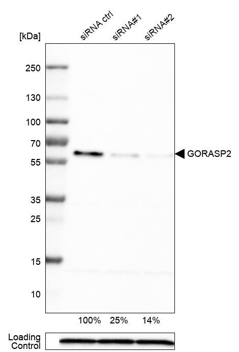
- Main image

- Experimental details
- Western blot analysis in U-251MG cells transfected with control siRNA, target specific siRNA probe #1 and #2, using Anti-GORASP2 antibody. Remaining relative intensity is presented. Loading control: Anti-PPIB.
- Sample type
- Human
- Protocol
- Protocol
Supportive validation
- Submitted by
- Atlas Antibodies (provider)
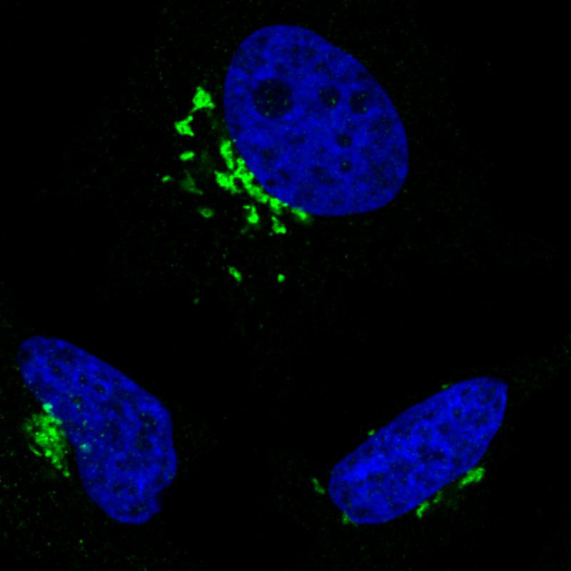
- Main image
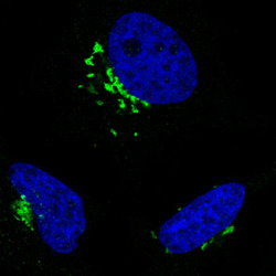
- Experimental details
- Immunofluorescence staining in HeLa cell line with Anti-GORASP2 monoclonal antibody, showing specific staining of the golgi apparatus in green. Microtubule- and nuclear probes are visualized in red and blue respectively (where available).
- Sample type
- Human
Supportive validation
- Submitted by
- Atlas Antibodies (provider)
- Enhanced method
- Orthogonal validation
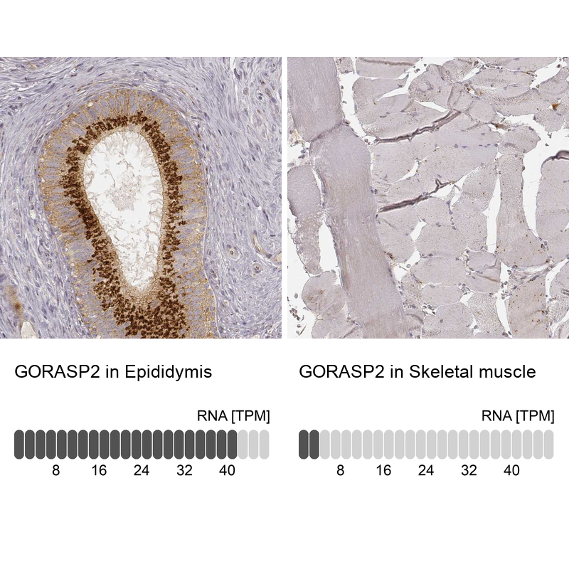
- Main image
- Experimental details
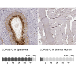
- Immunohistochemistry analysis in human epididymis and skeletal muscle tissues using AMAb91016 antibody. Corresponding GORASP2 RNA-seq data are presented for the same tissues.
- Sample type
- Human
- Protocol
- Protocol