 Explore
 Validate
 Learn
Learn Western blot
Antibody data
- Antibody Data
- Antigen structure
- References [0]
- Comments [0]
- Validations
- Western blot [1]
- ELISA [1]
- Immunohistochemistry [1]
Submit
Validation data
Reference
Comment
Report error
- Product number
- 23750002-0.1mg - Provider product page

- Provider
- Novus Biologicals
- Product name
- Rabbit Polyclonal IMP2/IGF2BP2 Antibody
- Antibody type
- Polyclonal
- Description
- Immunogen affinity purified. This product is specific for Human IGF2BP2.
- Reactivity
- Human
- Host
- Rabbit
- Isotype
- IgG
- Vial size
- 0.1 mg
- Storage
- Store at 4C short term. Aliquot and store at -20C long term. Avoid freeze-thaw cycles.
No comments: Submit comment
Supportive validation
- Submitted by
- Novus Biologicals (provider)
- Main image
- Experimental details
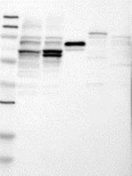
- Western Blot: IMP2/IGF2BP2 Antibody [23750002] - Samples: Lane 1, Marker [kDa]: 250, 130, 95, 72, 55, 36, 28, 17, 11 Lane 2, RT-4 Lane 3, U-251MG sp Lane 4, Human Plasma Lane 5, Liver Lane 6, Tonsil Target weight [kDa]: 66, 62 (splice variants) Validation score: 2 Validation description: Supportive - Band of predicted size in kDa (+/-20%) with additional bands present.

Supportive validation
- Submitted by
- Novus Biologicals (provider)
- Main image
- Experimental details
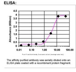
- ELISA: IMP2/IGF2BP2 Antibody [23750002]

Supportive validation
- Submitted by
- Novus Biologicals (provider)
- Main image

- Experimental details
- Immunohistochemistry: IMP2/IGF2BP2 Antibody [23750002] - Both normal and malignant tissues showed moderate to strong cytoplasmic staining. In normal tissues, surface epithelial cells of bronchus, glandular cells of the intestine, endometrium and cervix, and trophoblastic cells displayed strong cytoplasmic staining. Pancreas, surface epithelial cells of oral mucosa, vagina and cervix, non-neuronal cells of CNS tissues and lymphoid tissues were negative. Colo-rectal, skin and testis carcinomas exhibited strong cytoplasmic staining while ovarian, stomach and pancreas malignancies displayed moderate to strong cytoplasmic staining. Malignant carcinoids showed weak cytoplasmic and nuclear staining. Renal and prostate malignancies were weak to negative. Image and statement courtesy of the Human Protein Atlas (HPA).
