Explore
 Validate
 Learn
Learn Western blot
Antibody data
- Antibody Data
- Antigen structure
- References [0]
- Comments [0]
- Validations
- Western blot [3]
- Immunocytochemistry [1]
- Immunohistochemistry [5]
- Flow cytometry [2]
Submit
Validation data
Reference
Comment
Report error
- Product number
- TA501269 - Provider product page

- Provider
- Invitrogen Antibodies
- Product name
- IGF2BP2 Monoclonal Antibody (OTI3F9), TrueMAB™
- Antibody type
- Monoclonal
- Antigen
- Recombinant full-length protein
- Reactivity
- Human, Canine
- Host
- Mouse
- Isotype
- IgG
- Antibody clone number
- OTI3F9
- Vial size
- 100 µL
- Concentration
- 0.6 mg/mL
- Storage
- -20° C, Avoid Freeze/Thaw Cycles
No comments: Submit comment
Supportive validation
- Submitted by
- Invitrogen Antibodies (provider)
- Main image
- Experimental details
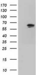
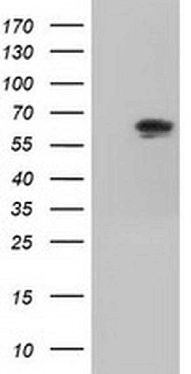
- HEK293T cells were transfected with the pCMV6-ENTRY control (Left lane) or pCMV6-ENTRY IGF2BP2 (RC205673, Right lane) cDNA for 48 hrs and lysed. Equivalent amounts of cell lysates (5 µg per lane) were separated by SDS-PAGE and immunoblotted with anti-IGF2. Positive lysates LY401961 (100 µg) and LC401961 (20 µg) can be purchased separately from OriGene.
- Submitted by
- Invitrogen Antibodies (provider)
- Main image
- Experimental details
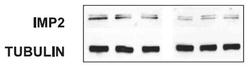
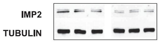
- Figure from citation: Western Blot of IGF2BP2(IMP2) protein level by using anti-IGF2BP2 antibody in rat glomerular MCs. View Citation
- Submitted by
- Invitrogen Antibodies (provider)
- Main image
- Experimental details
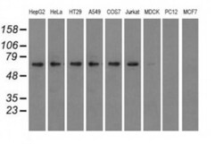
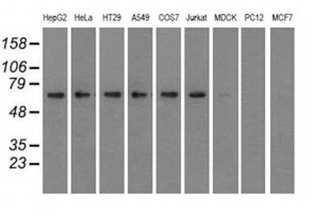
- Western blot analysis of extracts (35 µg) from 9 different cell lines by using anti-IGF2BP2 monoclonal antibody.
Supportive validation
- Submitted by
- Invitrogen Antibodies (provider)
- Main image
- Experimental details
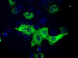
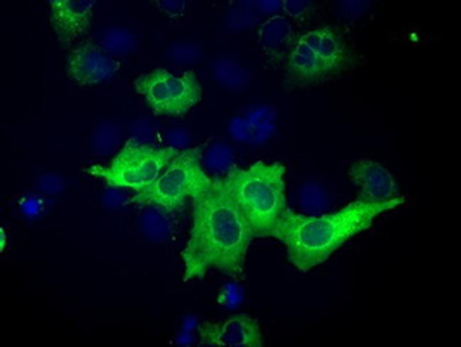
- Anti-IGF2BP2 mouse monoclonal antibody (TA501269) immunofluorescent staining of COS7 cells transiently transfected by pCMV6-ENTRY IGF2BP2(RC205673).
Supportive validation
- Submitted by
- Invitrogen Antibodies (provider)
- Main image
- Experimental details
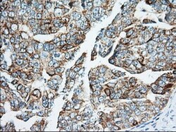
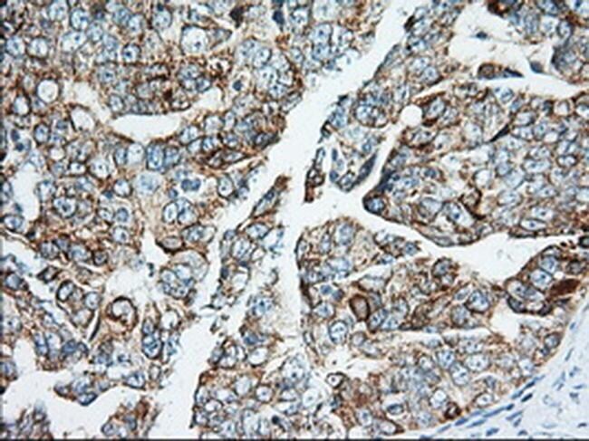
- Immunohistochemical staining of paraffin-embedded Adenocarcinoma of Human ovary tissue using anti-IGF2BP2 mouse monoclonal antibody. (Heat-induced epitope retrieval by 10mM citric buffer, pH6.0, 100°C for 10min, TA501269, Dilution 1:50)
- Submitted by
- Invitrogen Antibodies (provider)
- Main image
- Experimental details
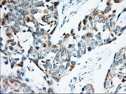
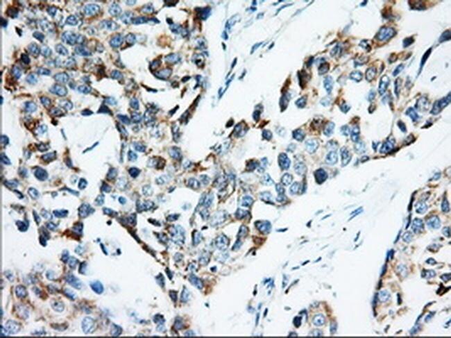
- Immunohistochemical staining of paraffin-embedded Adenocarcinoma of Human breast tissue using anti-IGF2BP2 mouse monoclonal antibody. (Heat-induced epitope retrieval by 10mM citric buffer, pH6.0, 100°C for 10min, TA501269, Dilution 1:50)
- Submitted by
- Invitrogen Antibodies (provider)
- Main image
- Experimental details
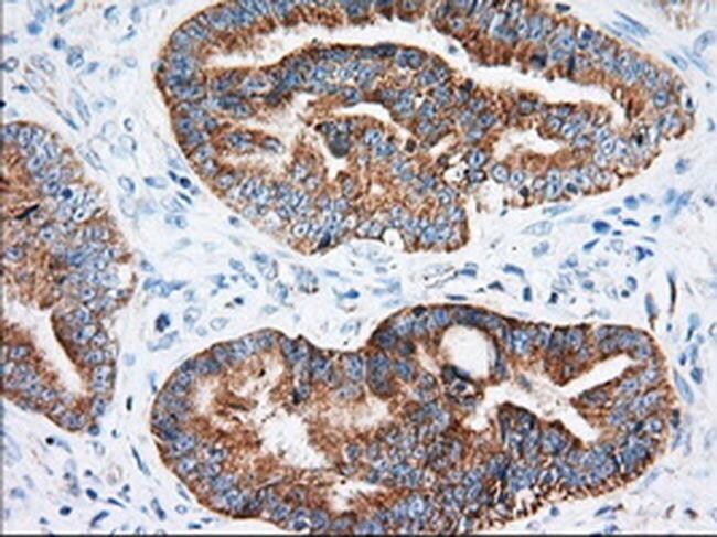
- Immunohistochemical staining of paraffin-embedded Adenocarcinoma of Human colon tissue using anti-IGF2BP2 mouse monoclonal antibody. (Heat-induced epitope retrieval by 10mM citric buffer, pH6.0, 100°C for 10min, TA501269, Dilution 1:50)
- Submitted by
- Invitrogen Antibodies (provider)
- Main image
- Experimental details
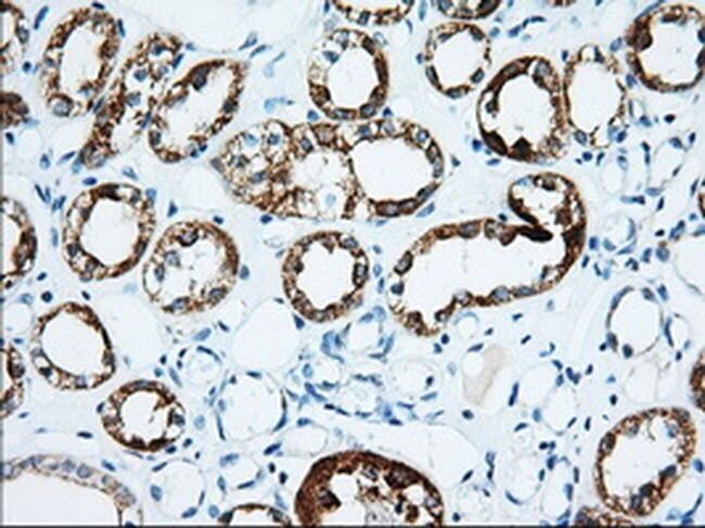
- Immunohistochemical staining of paraffin-embedded human Kidney tissue within the normal limits using anti-IGF2BP2 mouse monoclonal antibody. (Heat-induced epitope retrieval by 10mM citric buffer, pH6.0, 100°C for 10min, TA501269, Dilution 1:50)
- Submitted by
- Invitrogen Antibodies (provider)
- Main image
- Experimental details
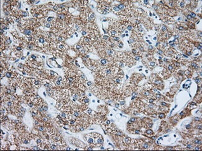
- Immunohistochemical staining of paraffin-embedded human liver tissue within the normal limits using anti-IGF2BP2 mouse monoclonal antibody. (Heat-induced epitope retrieval by 10mM citric buffer, pH6.0, 100°C for 10min, TA501269, Dilution 1:50)
Supportive validation
- Submitted by
- Invitrogen Antibodies (provider)
- Main image
- Experimental details
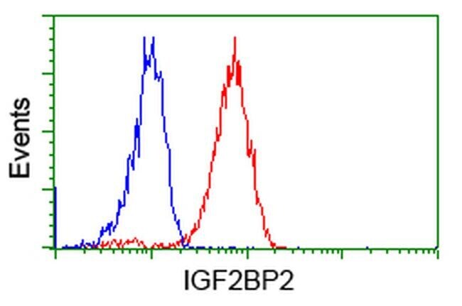
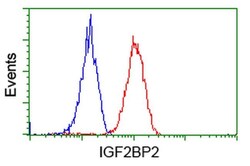
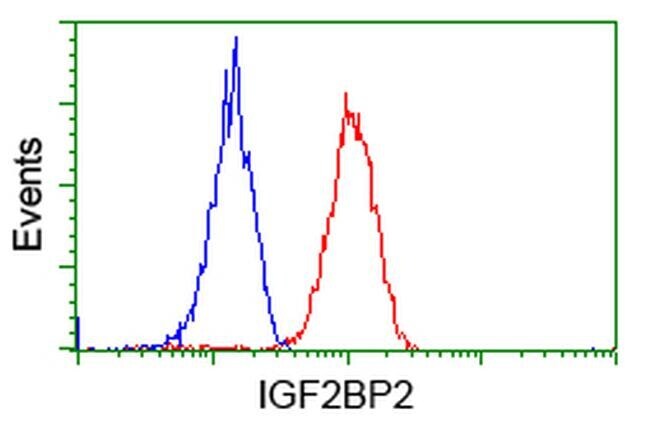
- Flow cytometric analysis of Hela cells, using anti-IGF2BP2 antibody (TA501269), (Red), compared to a nonspecific negative control antibody (TA50011), (Blue).
- Submitted by
- Invitrogen Antibodies (provider)
- Main image
- Experimental details
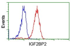
- Flow cytometric analysis of Jurkat cells, using anti-IGF2BP2 antibody (TA501269), (Red), compared to a nonspecific negative control antibody (TA50011), (Blue).