Explore
 Validate
 Learn
Learn Western blot
 Immunohistochemistry
Antibody data
- Antibody Data
- Antigen structure
- References [2]
- Comments [0]
- Validations
- Immunohistochemistry [1]
- Other assay [7]
Submit
Validation data
Reference
Comment
Report error
- Product number
- PA5-28899 - Provider product page

- Provider
- Invitrogen Antibodies
- Product name
- DLX1 Polyclonal Antibody
- Antibody type
- Polyclonal
- Antigen
- Recombinant full-length protein
- Description
- Recommended positive controls: U87-MG, mouse brain, rat brain. Predicted reactivity: Mouse (98%), Rat (98%), Chicken (89%), Rhesus Monkey (100%), Bovine (98%). Store product as a concentrated solution. Centrifuge briefly prior to opening the vial.
- Reactivity
- Human, Mouse, Rat
- Host
- Rabbit
- Isotype
- IgG
- Vial size
- 100 μL
- Concentration
- 1 mg/mL
- Storage
- Store at 4°C short term. For long term storage, store at -20°C, avoiding freeze/thaw cycles.
Submitted references Transcriptional network involving ERG and AR orchestrates Distal-less homeobox-1 mediated prostate cancer progression.
DLX1 acts as a crucial target of FOXM1 to promote ovarian cancer aggressiveness by enhancing TGF-β/SMAD4 signaling.
Goel S, Bhatia V, Kundu S, Biswas T, Carskadon S, Gupta N, Asim M, Morrissey C, Palanisamy N, Ateeq B
Nature communications 2021 Sep 7;12(1):5325
Nature communications 2021 Sep 7;12(1):5325
DLX1 acts as a crucial target of FOXM1 to promote ovarian cancer aggressiveness by enhancing TGF-β/SMAD4 signaling.
Chan DW, Hui WW, Wang JJ, Yung MM, Hui LM, Qin Y, Liang RR, Leung TH, Xu D, Chan KK, Yao KM, Tsang BK, Ngan HY
Oncogene 2017 Mar;36(10):1404-1416
Oncogene 2017 Mar;36(10):1404-1416
No comments: Submit comment
Supportive validation
- Submitted by
- Invitrogen Antibodies (provider)
- Main image
- Experimental details
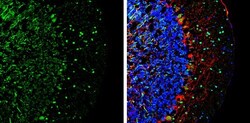
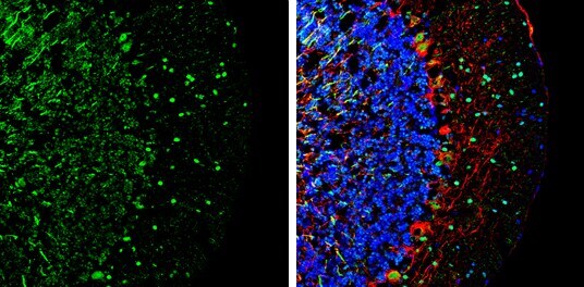
- Immunohistochemistry (Frozen) analysis of DLX1 was performed in frozen-sectioned mouse cerebellum tissue using DLX1 Polyclonal Antibody (Product # PA5-28899) at a dilution of 1:250 (Green). Red: NF-H, stained by NF-H antibody diluted at 1:500. Blue: Fluoroshield with DAPI.
Supportive validation
- Submitted by
- Invitrogen Antibodies (provider)
- Main image
- Experimental details
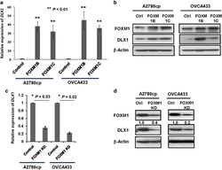
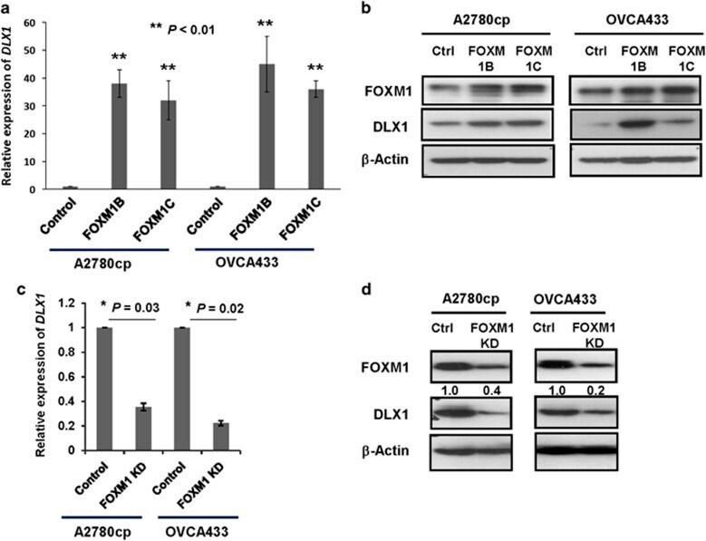
- Figure 2 Identification of DLX1 as a downstream target of FOXM1. ( a ) The qRT-PCR analysis showed that DLX1 expression was upregulated by 40-fold and 35-fold in the A2780cp cells, and by 50-fold and 40-fold in OVCA433 cells (** P
- Submitted by
- Invitrogen Antibodies (provider)
- Main image

- Experimental details
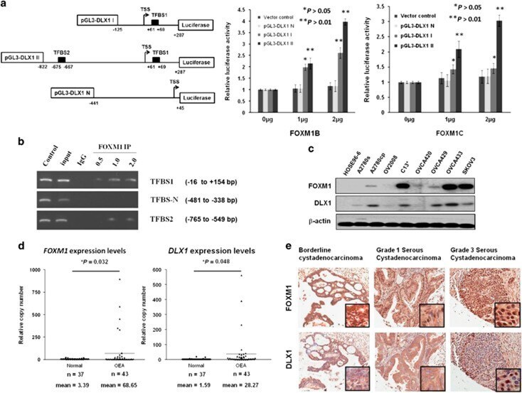
- Figure 3 FOXM1 and DLX1 are concomitantly expressed in high-grade serous ovarian cancers. ( a ) TFBS1 and TFBS2 are the two conserved FOXM1-binding sites on the DLX1 promoter. (left) A schematic diagram illustrating the DLX1 promoter luciferase reporter constructs: pGL3-DLX1 I with only one putative FOXM1-binding site, pGL3-DLX1 II with both putative FOXM1-binding sites, and pGL3-DLX1 II with none of the predicted binding sites. (right) The luciferase promoter assay demonstrated the ability of FOXM1 to transcriptionally activate DLX1 by binding to the two FOXM1-binding sites. Both FOMX1B and FOXM1C could increase the luciferase activity of the pGL3-DLX1 I and pGL3-DLX1 II constructs in a concentration-dependent manner (* P
- Submitted by
- Invitrogen Antibodies (provider)
- Main image
- Experimental details
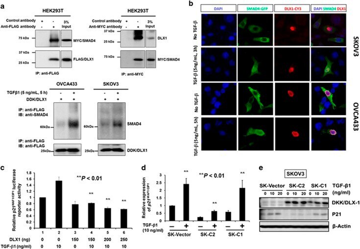
- Figure 6 DLX1 activates TGF-beta1/SMAD4 signaling through a direct interaction with SMAD4 in the nuclei of ovarian cancer cells. ( a ) Reciprocal co-immunoprecipitation (co-IP) demonstrated that DLX1 interacts with SMAD4. (upper) Co-transfection of DDK/MYC/DLX1 and MYC/SMAD4 in HEK293T cells and co-IP using anti-Flag (DDK) showed that DDK/MYC/DLX1 could pull down MYC/SMAD4 confirmed by immunoblotting using anti-MYC. Conversely, transfection of MYC/SMAD4 in HEK293T cells showed that co-IP using anti-MYC could pull down endogenous DLX1 confirmed by immunoblotting using anti-DLX1. (lower) Co-IP assay using anti-Flag (DDK) confirmed that TGF-beta1 (5 ng/ml, 5 h) induction could enhance DDK/MYC/DLX1 to pull down more MYC/SMAD4 in SKOV3 and OVCA433 cells, indicating that TGF-beta1 induces more MYC/SMAD4 translocating into the nucleus and increases DLX1-SMAD4 interaction. ( b ) Immunofluorescent analysis demonstrating DLX1-SMAD4 co-localization in the nuclei of the SKOV3 and OVCA433 ovarian cancer cells after TGF-beta1 (5 ng/ml, 3 h and 5 h for SKOV3 and OVCA433, respectively) stimulation. ( c ) DLX1 inhibits p21 WAF1/CIP1 promoter activity upon TGF-beta1 treatment. Various amounts of the Myc-DDK-tagged HuDLX1 vector were co-transfected with the pWWP-luciferase reporter construct and empty vector pCMV2 into HEK293 cells. TGF-beta1 (10 ng/ml) was added 24 h after transfection and incubated for another 5 h before the luciferase activity was measured. The relative p21 WAF1/CIP1 lucifer
- Submitted by
- Invitrogen Antibodies (provider)
- Main image
- Experimental details
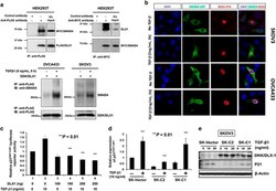
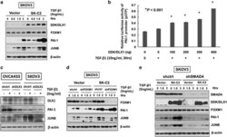
- Figure 7 DLX1 is a critical downstream effector of FOXM1 that activates TGF-beta1/SMAD4 signaling. ( a ) The western blot analysis showed increased expression of two TGF-beta signaling targets, PAI-1 and JUNB, in SKOV3 cells stably expressing DLX1 (SK-C2) in response to TGF-beta1 (5 ng/ml) treatment compared with the vector control (SK-vector). ( b ) DLX1 induced a concentration-dependent increase in PAI-1 luciferase activity upon TGF-beta1 treatment (10 ng/ml, 3 h). ( c ) Depletion of DLX1 by lentiviral shRNA approach remarkably reduce the expressions of PAI-1 and JUNB in OVCA433 and SKOV3 cells upon treatment of TGF-beta1 (5 ng/ml) for 5 h. ( d ) FOXM1 knockdown does not alter the ability of DLX1 to upregulate PAI-1 and JUNB expression upon TGF-beta1 (5 ng/ml) treatment in a SKOV3 cell model stably expressing DLX1 (SK-C2). ( e ) The western blot analysis showed that SMAD4 knockdown markedly attenuated TGFbeta1-induced expression of PAI-1 and JUNB, with or without stably overexpressing DDK/DLX1 in SKOV3 cells (SK-C2 and empty vector control).

- Submitted by
- Invitrogen Antibodies (provider)
- Main image
- Experimental details
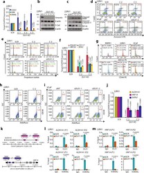
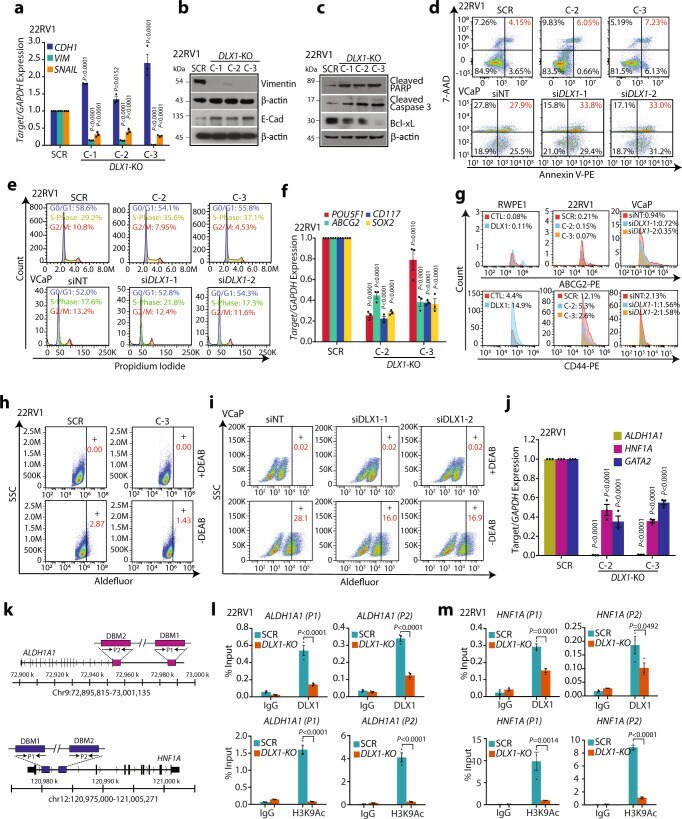
- Fig. 2 Genetic ablation of DLX1 inhibits oncogenic properties. a Q-PCR data showing expression of EMT markers in 22RV1- DLX1 -KO and control SCR cells. b Immunoblots showing vimentin and E-cadherin using same cells as a . beta-actin was used as loading control. c Same as b except for cleaved PARP, cleaved Caspase-3 and Bcl-xL. d Flow cytometry-based apoptosis assay using 22RV1- DLX1 -KO and control cells (top panel) and DLX1 -silenced VCaP cells (bottom panel). e Flow cytometry data for cell cycle distribution using same cells as in d . f Q-PCR data for stem cell markers using same cells as d . g Flow cytometry data depicting ABCG2 (top panel) and CD44 (bottom panel) expression in DLX1 overexpressing RWPE1 cells, 22RV1- DLX1 -KO, and DLX1 -silenced VCaP cells. h Fluorescence intensity of catalyzed ALDH substrate in 22RV1- DLX1 -KO and control cells. Marked windows show ALDH1 + percent cell population. i Same as h , except for DLX1 silenced VCaP cells. j Q-PCR data showing expression of target genes in 22RV1- DLX1 -KO and control cells( P < 0.0001). k Schema showing the chromosomal location of DLX1 binding motif (DBM1/2) at the ALDLH1A1 (top) and HNF1A (bottom) promoters. l ChIP-qPCR data of DLX1 (top panel) and H3K9Ac (bottom panel) on ALDH1A1 in 22RV1- DLX1 -KO and SCR control cells ( P < 0.0001). m Same as in l except for the HNF1A promoter. Data shown from three biologically independent samples ( n = 3). Data represent mean +- SEM. For panels, a , f , and j Two-way ANOVA,
- Submitted by
- Invitrogen Antibodies (provider)
- Main image
- Experimental details
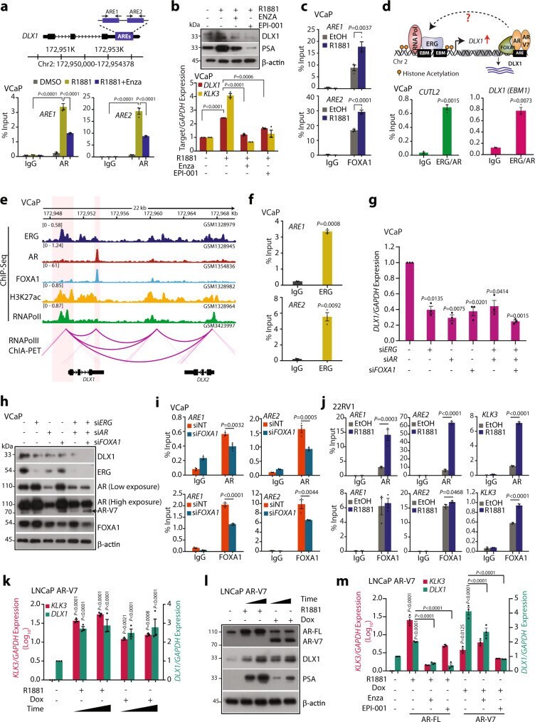
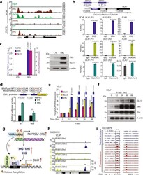
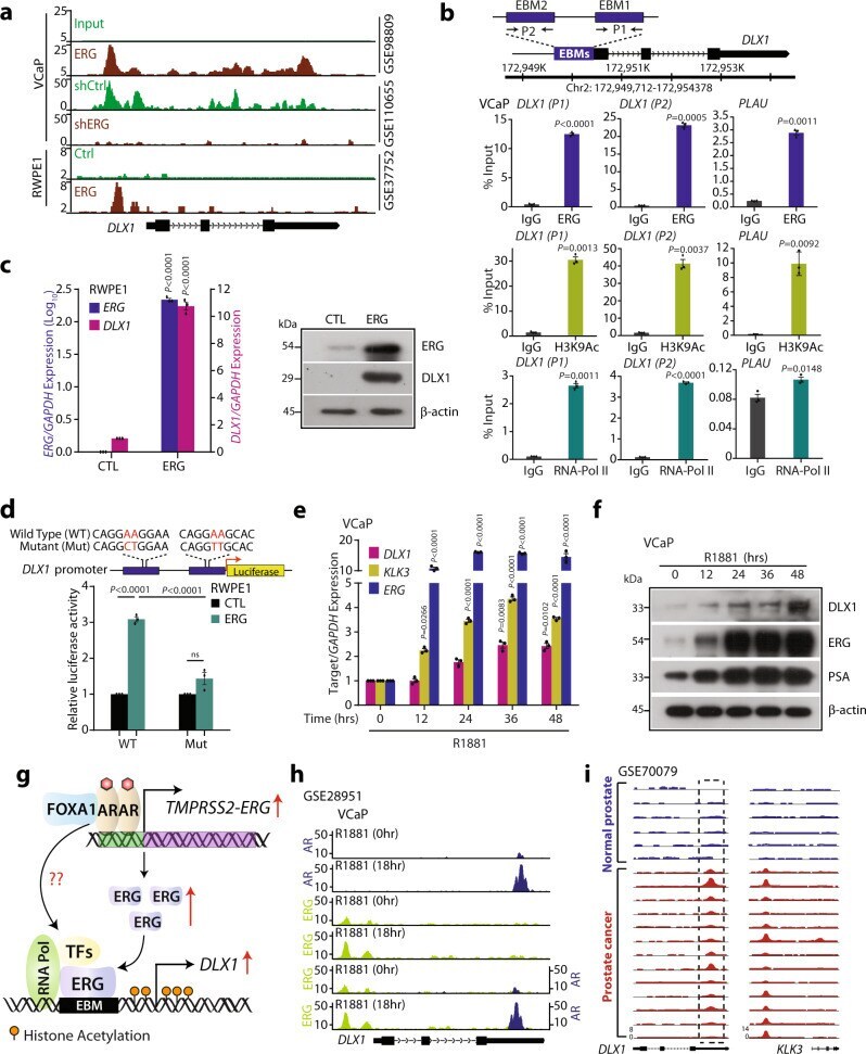
- Fig. 5 DLX1 is a transcriptional target of ERG in TMPRSS2-ERG positive prostate cancer. a Chromatin immunoprecipitation-sequencing (ChIP-Seq) data depicting ERG enrichment at the DLX1 promoter in the indicated cell lines from GEO databases (GSE98809, GSE110655, and GSE37752). b Schema showing chromosomal location of the ERG binding motifs (EBM) onto DLX1 promoter selected for ChIP-qPCR (top panel). ChIP-qPCR data showing recruitment of ERG, histone H3 lysine 9 acetylation (H3K9Ac), and RNA-polymerase II (RNA-Pol II) at the DLX1 and PLAU promoters. c Q-PCR (left panel) and immunoblot (right panel) data showing the expression of ERG and DLX1 in RWPE1 cells overexpressing ERG ( P < 0.0001). d Schema showing site-directed mutagenesis of DLX1 promoter cloned upstream of luciferase gene, nucleotides in red were mutated (top). Luciferase reporter assay indicating wild-type (WT) and mutant (Mut) DLX1 promoter-driven reporter activity (bottom panel) in ERG overexpressing and control RWPE1 cells ( P < 0.0001). e Q-PCR data showing relative expression of target genes in VCaP cells stimulated with 10 nM R1881 at the indicated time points. f Same as e , except immunoblot data. g Schematic diagram depicting ERG-mediated transcriptional regulation of DLX1 and plausible role of AR in DLX1 regulation. h ChIP-Seq data (GSE28951) showing recruitment of AR and ERG on the DLX1 enhancer and promoter regions respectively, in R1881-stimulated VCaP cells. i ChIP-Seq data (GSE70079) showing enrichment
- Submitted by
- Invitrogen Antibodies (provider)
- Main image
- Experimental details
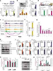
- Fig. 6 AR and AR-V7 regulate DLX1 expression in both ERG-dependent and -independent manners. a Schematic showing the androgen response elements (AREs) at the DLX1 putative enhancer (top panel). ChIP-qPCR data (bottom panel) depicting AR recruitment at the DLX1 putative enhancer in R1881 (10 nM) stimulated VCaP cells in the presence or absence of Enzalutamide (Enza, 10 uM) ( P < 0.0001). b Immunoblot (top panel) and Q-PCR (bottom panel) data showing relative expression of target genes in VCaP cells under similar culture conditions as indicated. c ChIP-qPCR data depicting FOXA1 recruitment at the DLX1 putative enhancer in R1881 (10 nM) stimulated VCaP. d Schematic representation showing the possible interaction between ERG and AR on the DLX1 promoter (top panel). Re-ChIP data showing co-enrichment of AR and ERG on EBM at the DLX1 promoter (bottom panel). e Integrated genome view of 3D-chromatin structure and binding of transcription factors at the genomic and nearby region of DLX1 . f Bar plots depicting ChIP-qPCR data for ERG occupancy at the DLX1 enhancer region. g Q-PCR data showing relative expression of DLX1 in siRNA-mediated ERG , AR, and/or FOXA1 -silenced VCaP cells. h Same as g except immunoblot data. i ChIP-qPCR data showing enrichment of AR (top panel) and FOXA1 (bottom panel) in siRNA-mediated FOXA1 -silenced VCaP cells . j ChIP-qPCR data depicting AR (top panel) and FOXA1 (bottom panel) enrichment at the DLX1 putative enhancer in 22RV1 cells stimulated with R1881 (