Explore
 Validate
 Learn
Learn Immunohistochemistry
Antibody data
- Antibody Data
- Antigen structure
- References [0]
- Comments [0]
- Validations
- Immunohistochemistry [1]
- Flow cytometry [1]
Submit
Validation data
Reference
Comment
Report error
- Product number
- MAB65761-100 - Provider product page

- Provider
- Novus Biologicals
- Product name
- Mouse Monoclonal B7-H4 Antibody
- Antibody type
- Monoclonal
- Description
- Protein A or G purified from hybridoma culture supernatant. Detects human B7-H4 in direct ELISAs.
- Reactivity
- Human
- Host
- Mouse
- Conjugate
- Unconjugated
- Isotype
- IgG
- Vial size
- 100 ug
- Storage
- Use a manual defrost freezer and avoid repeated freeze-thaw cycles. 12 months from date of receipt, -20 to -70 degreesC as supplied. 1 month, 2 to 8 degreesC under sterile conditions after reconstitution. 6 months, -20 to -70 degreesC under sterile conditions after reconstitution.
No comments: Submit comment
Supportive validation
- Submitted by
- Novus Biologicals (provider)
- Main image

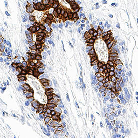
- Experimental details
- B7-H4 in Human Breast. B7-H4 was detected in immersion fixed paraffin-embedded sections of human breast using Mouse Anti-Human B7-H4 Monoclonal Antibody (Catalog # MAB65761) at 1.7 µg/mL for 1 hour at room temperature followed by incubation with the Anti-Mouse IgG VisUCyte™ HRP Polymer Antibody (Catalog # VC001). Tissue was stained using DAB (brown) and counterstained with hematoxylin (blue). Specific staining was localized to plasma membrane in epithelial cells. View our protocol for IHC Staining with VisUCyte HRP Polymer Detection Reagents.
Supportive validation
- Submitted by
- Novus Biologicals (provider)
- Main image
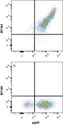
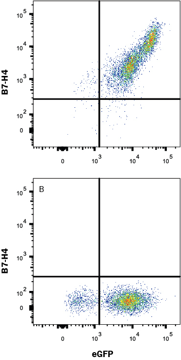
- Experimental details
- Detection of B7-H4 in HEK293 Human Cell Line Transfected with Human B7-H4 and eGFP by Flow Cytometry. HEK293 human embryonic kidney cell line transfected with either (A) human B7-H4 or (B) irrelevant transfectants and eGFP were stained with Mouse Anti-Human B7-H4 Monoclonal Antibody (Catalog # MAB65761) followed by Allophycocyanin-conjugated Anti-Mouse IgG Secondary Antibody (Catalog # F0101B). View our protocol for Staining Membrane-associated Proteins.