Explore
 Validate
 Learn
Learn Western blot
Antibody data
- Antibody Data
- Antigen structure
- References [0]
- Comments [0]
- Validations
- Western blot [2]
- Immunohistochemistry [1]
- Flow cytometry [1]
Submit
Validation data
Reference
Comment
Report error
- Product number
- 10-4172-25 - Provider product page

- Provider
- ABEOMICS Inc.
- Product name
- Anti-B7-H4 Antibody
- Antibody type
- Monoclonal
- Description
- B7-H4, a member of B7 family, is a transmembrane protein that has been shown to inhibit T cell responses and neutrophil expansion during bacterial infections. B7-H4 mRNA is widely distributed in mouse and human peripheral tissues and can be induced in monocytes, macrophages, and dendritic cells upon IL-6 and IL-10 stimulation. However, in a variety of tumor cells, B7-H4 is predominantly present in intracellular compartments with unknown mechanism and functions. Cell surface expression of B7-H4 protein is limited and shows an inducible pattern on hematopoietic cells. The overexpression of B7-H4 has been found in various types of human tumors, such as breast cancer, renal cell carcinoma (RCC), ovarian cancer, esophageal squamous cell carcinoma, gastric cancer, pancreatic cancer and melanoma etc, where its expression level is positively correlated with disease progression. By arresting cell cycle, B7-H4 ligation of T cells has a profound inhibitory effect on the growth, cytokine secretion, and development of cytotoxicity.
- Reactivity
- Human
- Host
- Mouse
- Conjugate
- Unconjugated
- Antigen sequence
A partial length recombinant protei
n  (a.a 23-220) of B7-H4 was used a
s the immunogen for this antibody.- Isotype
- IgG
- Antibody clone number
- ABM53A6
- Vial size
- 100 µg
- Concentration
- 0.5 mg/ml
- Storage
- Store the antibody at 4°C, stable for 6 months. For long-term storage, store at -20°C. Avoid repeat freez thawing
No comments: Submit comment
Supportive validation
- Submitted by
- ABEOMICS Inc. (provider)
- Main image
- Experimental details
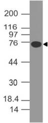
- Western blot analysis of B7-H4. Anti-B7-H4 antibody (Clone: ABM53A6) was used at 4 µg/ml on human B7H4-FC fusion protein Lysate.
- Protocol
- Protocol

- Submitted by
- ABEOMICS Inc. (provider)
- Main image

- Experimental details
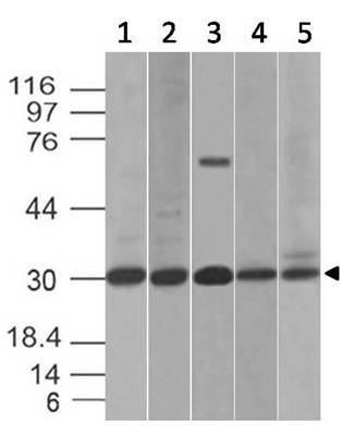
- Western blot analysis of B7-H4. Anti-B7-H4 antibody (Clone: ABM53A6) was used at 2 µg/ml on (1) HCT-116, (2) PC3, (3) Kato 111, (4) C2C12 and (5) RAW Lysates.
- Protocol
- Protocol
Supportive validation
- Submitted by
- ABEOMICS Inc. (provider)
- Main image

- Experimental details
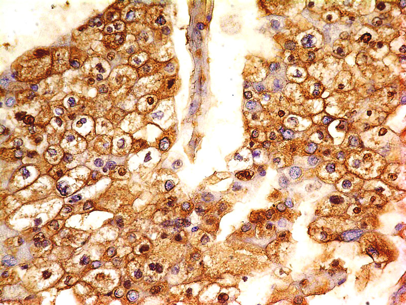
- Immunohistochemical analysis of B7-H4 antibody in Renal cell carcinoma using anti-B7-H4 antibody (Clone: ABM53A6) at 5 μg/ml
- Protocol
- Protocol
Supportive validation
- Submitted by
- ABEOMICS Inc. (provider)
- Main image
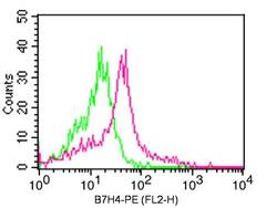
- Experimental details
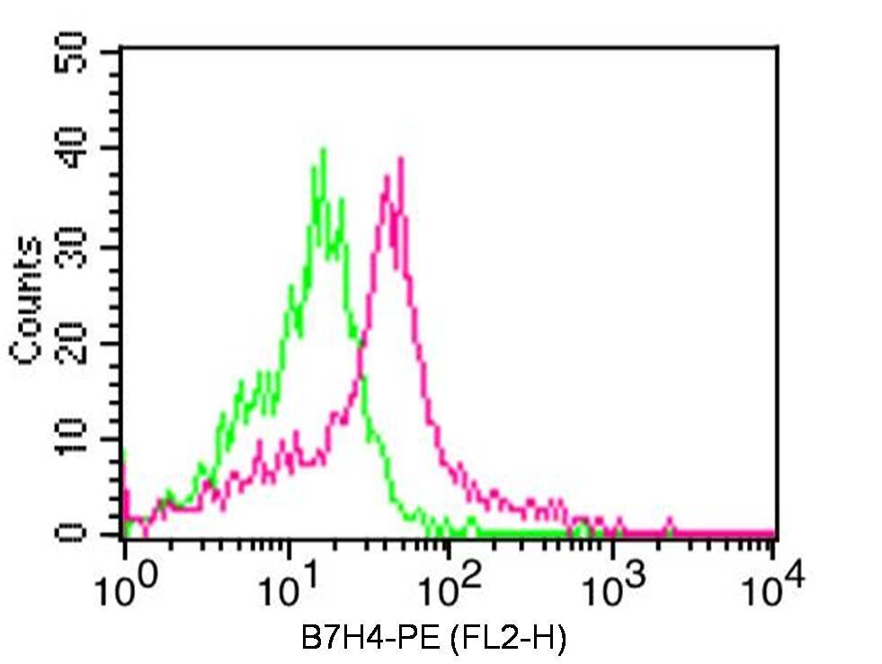
- Cell surface Flow analysis of B7-H4 antibody in human PBMC (Monocytes) cells using 0.5 μg/ 10^6 cells of anti-B7-H4 antibody (ABM53A6). Green represents isotype control;red represents anti-B7H4 antibody. Goat anti-mouse PE conjugate was used as secondary antibody.
- Protocol
- Protocol