Explore
 Validate
 Learn
Learn Western blot
 ELISA
ELISAAntibody data
- Antibody Data
- Antigen structure
- References [8]
- Comments [0]
- Validations
- Western blot [2]
- Immunohistochemistry [2]
Submit
Validation data
Reference
Comment
Report error
- Product number
- H00003283-M01 - Provider product page

- Provider
- Novus Biologicals
- Proper citation
- Novus Cat#H00003283-M01, RRID:AB_539678
- Product name
- Mouse Monoclonal HSD3B1 Antibody
- Antibody type
- Monoclonal
- Description
- IgG purified. HSD3B1 - hydroxy-delta-5-steroid dehydrogenase, 3 beta- and steroid delta-isomerase 1
- Reactivity
- Human
- Host
- Mouse
- Isotype
- IgG
- Vial size
- 0.1 mg
- Storage
- Aliquot and store at -20C or -80C. Avoid freeze-thaw cycles.
Submitted references Lineage-Specific Alterations in Gynecologic Neoplasms with Choriocarcinomatous Differentiation: Implications for Origin and Therapeutics.
A case of primary aldosteronism caused by unilateral multiple adrenocortical micronodules presenting as muscle cramps at rest: The importance of functional histopathology for identifying a culprit lesion.
Expression of steroidogenic enzymes and their transcription factors in cortisol-producing adrenocortical adenomas: immunohistochemical analysis and quantitative real-time polymerase chain reaction studies.
Steroidogenic enzymes, their related transcription factors and nuclear receptors in human sebaceous glands under normal and pathological conditions.
Chorangiocarcinoma: a case report and review of the literature.
Advances in the diagnosis of gestational trophoblastic tumors and tumor-like lesions.
HSD3B1 as a novel trophoblast-associated marker that assists in the differential diagnosis of trophoblastic tumors and tumorlike lesions.
Trophogram, an immunohistochemistry-based algorithmic approach, in the differential diagnosis of trophoblastic tumors and tumorlike lesions.
Xing D, Zheng G, Pallavajjala A, Schoolmeester JK, Liu Y, Haley L, Hu Y, Liu L, Logan L, Lin Y, Pearce KE, Sattler CA, Tsai YC, Vang R, Hung CF, Wu TC, Ronnett BM
Clinical cancer research : an official journal of the American Association for Cancer Research 2019 Jul 15;25(14):4516-4529
Clinical cancer research : an official journal of the American Association for Cancer Research 2019 Jul 15;25(14):4516-4529
A case of primary aldosteronism caused by unilateral multiple adrenocortical micronodules presenting as muscle cramps at rest: The importance of functional histopathology for identifying a culprit lesion.
Ito A, Yamazaki Y, Sasano H, Matsubara D, Fukushima N, Tamba M, Tabata K, Ashizawa K, Takei A, Koizumi M, Sakuma Y, Sata N, Oshiro H
Pathology international 2017 Apr;67(4):214-221
Pathology international 2017 Apr;67(4):214-221
Expression of steroidogenic enzymes and their transcription factors in cortisol-producing adrenocortical adenomas: immunohistochemical analysis and quantitative real-time polymerase chain reaction studies.
Kubota-Nakayama F, Nakamura Y, Konosu-Fukaya S, Azmahani A, Ise K, Yamazaki Y, Kitawaki Y, Felizola SJ, Ono Y, Omata K, Morimoto R, Iwama N, Satoh F, Sasano H
Human pathology 2016 Aug;54:165-73
Human pathology 2016 Aug;54:165-73
Steroidogenic enzymes, their related transcription factors and nuclear receptors in human sebaceous glands under normal and pathological conditions.
Azmahani A, Nakamura Y, Felizola SJ, Ozawa Y, Ise K, Inoue T, McNamara KM, Doi M, Okamura H, Zouboulis CC, Aiba S, Sasano H
The Journal of steroid biochemistry and molecular biology 2014 Oct;144 Pt B:268-79
The Journal of steroid biochemistry and molecular biology 2014 Oct;144 Pt B:268-79
Chorangiocarcinoma: a case report and review of the literature.
Ariel I, Boldes R, Weintraub A, Reinus C, Beller U, Arbel R
International journal of gynecological pathology : official journal of the International Society of Gynecological Pathologists 2009 May;28(3):267-71
International journal of gynecological pathology : official journal of the International Society of Gynecological Pathologists 2009 May;28(3):267-71
Advances in the diagnosis of gestational trophoblastic tumors and tumor-like lesions.
Mao TL, Shih IeM
Expert opinion on medical diagnostics 2009 Jul;3(4):371-80
Expert opinion on medical diagnostics 2009 Jul;3(4):371-80
HSD3B1 as a novel trophoblast-associated marker that assists in the differential diagnosis of trophoblastic tumors and tumorlike lesions.
Mao TL, Kurman RJ, Jeng YM, Huang W, Shih IeM
The American journal of surgical pathology 2008 Feb;32(2):236-42
The American journal of surgical pathology 2008 Feb;32(2):236-42
Trophogram, an immunohistochemistry-based algorithmic approach, in the differential diagnosis of trophoblastic tumors and tumorlike lesions.
Shih IeM
Annals of diagnostic pathology 2007 Jun;11(3):228-34
Annals of diagnostic pathology 2007 Jun;11(3):228-34
No comments: Submit comment
Supportive validation
- Submitted by
- Novus Biologicals (provider)
- Main image
- Experimental details
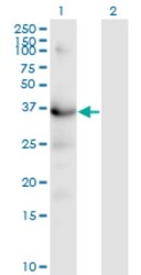
- Western Blot: HSD3B1 Antibody (3C11-D4) [H00003283-M01] - Analysis of HSD3B1 expression in transfected 293T cell line by HSD3B1 monoclonal antibody (M01), clone 3C11-D4.Lane 1: HSD3B1 transfected lysate (Predicted MW: 42.3 KDa).Lane 2: Non-transfected lysate.
- Submitted by
- Novus Biologicals (provider)
- Main image
- Experimental details
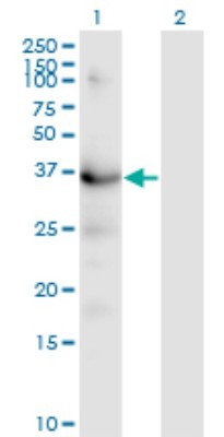
- Western Blot: HSD3B1 Antibody (3C11-D4) [H00003283-M01] - HSD3B1 monoclonal antibody (M01), clone 3C11-D4. Analysis of HSD3B1 expression in human placenta.

Supportive validation
- Submitted by
- Novus Biologicals (provider)
- Main image

- Experimental details
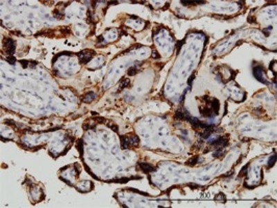
- Immunohistochemistry-Paraffin: HSD3B1 Antibody (3C11-D4) [H00003283-M01] - Analysis of monoclonal antibody to HSD3B1 on formalin-fixed paraffin-embedded human placenta. Antibody concentration 3 ug/ml.
- Submitted by
- Novus Biologicals (provider)
- Main image
- Experimental details
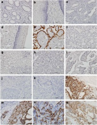
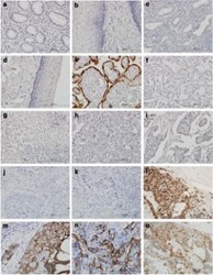
- Immunohistochemistry-Paraffin: HSD3B1 Antibody (3C11-D4) [H00003283-M01] - Immunohistochemical staining of various tissues with hematoxylin and HSD3B1 antibody under high magnification.Antibody concentration 3 ug/ml. ( a ) Stomach ( b ) Esophagus ( c ) Endometrium ( d ) Uterine cervix ( e ) Placenta ( f ) Ovary, clear cell carcinoma ( g ) Hepatocellular carcinoma ( h ) Breast cancer ( i ) Colon adenocarcinoma ( j and k ) Cervical carcinoma ( l, m, and n ) Choriocarcinoma ( o ) Epithelioid trophoblastic tumor.