Explore
 Validate
 Learn
Learn Western blot
 ELISA
ELISAAntibody data
- Antibody Data
- Antigen structure
- References [8]
- Comments [0]
- Validations
- Western blot [1]
- Immunocytochemistry [1]
- Immunoprecipitation [1]
- Immunohistochemistry [1]
Submit
Validation data
Reference
Comment
Report error
- Product number
- 11610-1-AP - Provider product page

- Provider
- Proteintech Group
- Proper citation
- Proteintech Cat#11610-1-AP, RRID:AB_10859665
- Product name
- MTX2 antibody
- Antibody type
- Polyclonal
- Description
- KD/KO validated MTX2 antibody (Cat. #11610-1-AP) is a rabbit polyclonal antibody that shows reactivity with human, mouse, rat and has been validated for the following applications: IF, IHC, IP, WB,ELISA.
- Reactivity
- Human, Mouse, Rat
- Host
- Rabbit
- Conjugate
- Unconjugated
- Isotype
- IgG
- Vial size
- 20ul, 150ul
Submitted references Disruption of axonal transport in Parkinson's disease: the role of pathological α-Syn and AMPK/p38 MAPK signaling.
Loss of MTX2 causes mitochondrial dysfunction, podocyte injury, nephrotic proteinuria and glomerulopathy in mice and patients.
Proteomic analysis implicates that postovulatory aging leads to aberrant gene expression, biosynthesis, RNA metabolism and cell cycle in mouse oocytes.
Loss of Sam50 in hepatocytes induces cardiolipin-dependent mitochondrial membrane remodeling to trigger mtDNA release and liver injury.
Mitochondrial cristae architecture protects against mtDNA release and inflammation.
Loss of MTX2 causes mandibuloacral dysplasia and links mitochondrial dysfunction to altered nuclear morphology.
Sam50-Mic19-Mic60 axis determines mitochondrial cristae architecture by mediating mitochondrial outer and inner membrane contact.
Sam50 Regulates PINK1-Parkin-Mediated Mitophagy by Controlling PINK1 Stability and Mitochondrial Morphology.
Yang X, Ma Z, Lian P, Wu Y, Liu K, Zhang Z, Tang Z, Xu Y, Cao X
NPJ Parkinson's disease 2025 May 6;11(1):114
NPJ Parkinson's disease 2025 May 6;11(1):114
Loss of MTX2 causes mitochondrial dysfunction, podocyte injury, nephrotic proteinuria and glomerulopathy in mice and patients.
Li T, Bao Y, Xia Y, Meng H, Zhou C, Huang L, Wang X, Lai EY, Jiang P, Mao J
International journal of biological sciences 2024;20(3):937-952
International journal of biological sciences 2024;20(3):937-952
Proteomic analysis implicates that postovulatory aging leads to aberrant gene expression, biosynthesis, RNA metabolism and cell cycle in mouse oocytes.
Zhang C, Dong X, Yuan X, Song J, Wang J, Liu B, Wu K
Journal of ovarian research 2022 Oct 14;15(1):112
Journal of ovarian research 2022 Oct 14;15(1):112
Loss of Sam50 in hepatocytes induces cardiolipin-dependent mitochondrial membrane remodeling to trigger mtDNA release and liver injury.
Chen L, Dong J, Liao S, Wang S, Wu Z, Zuo M, Liu B, Yan C, Chen Y, He H, Meng Q, Song Z
Hepatology (Baltimore, Md.) 2022 Nov;76(5):1389-1408
Hepatology (Baltimore, Md.) 2022 Nov;76(5):1389-1408
Mitochondrial cristae architecture protects against mtDNA release and inflammation.
He B, Yu H, Liu S, Wan H, Fu S, Liu S, Yang J, Zhang Z, Huang H, Li Q, Wang F, Jiang Z, Liu Q, Jiang H
Cell reports 2022 Dec 6;41(10):111774
Cell reports 2022 Dec 6;41(10):111774
Loss of MTX2 causes mandibuloacral dysplasia and links mitochondrial dysfunction to altered nuclear morphology.
Elouej S, Harhouri K, Le Mao M, Baujat G, Nampoothiri S, Kayserili H, Menabawy NA, Selim L, Paneque AL, Kubisch C, Lessel D, Rubinsztajn R, Charar C, Bartoli C, Airault C, Deleuze JF, Rötig A, Bauer P, Pereira C, Loh A, Escande-Beillard N, Muchir A, Martino L, Gruenbaum Y, Lee SH, Manivet P, Lenaers G, Reversade B, Lévy N, De Sandre-Giovannoli A
Nature communications 2020 Sep 11;11(1):4589
Nature communications 2020 Sep 11;11(1):4589
Sam50-Mic19-Mic60 axis determines mitochondrial cristae architecture by mediating mitochondrial outer and inner membrane contact.
Tang J, Zhang K, Dong J, Yan C, Hu C, Ji H, Chen L, Chen S, Zhao H, Song Z
Cell death and differentiation 2020 Jan;27(1):146-160
Cell death and differentiation 2020 Jan;27(1):146-160
Sam50 Regulates PINK1-Parkin-Mediated Mitophagy by Controlling PINK1 Stability and Mitochondrial Morphology.
Jian F, Chen D, Chen L, Yan C, Lu B, Zhu Y, Chen S, Shi A, Chan DC, Song Z
Cell reports 2018 Jun 5;23(10):2989-3005
Cell reports 2018 Jun 5;23(10):2989-3005
No comments: Submit comment
Supportive validation
- Submitted by
- Proteintech Group (provider)
- Main image

- Experimental details
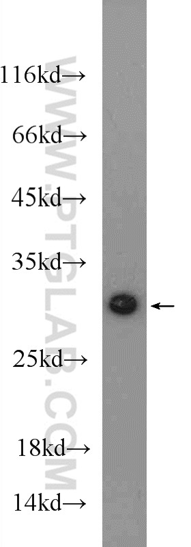
- mouse heart tissue were subjected to SDS PAGE followed by western blot with 11610-1-AP( MTX2 Antibody) at dilution of 1:600
- Sample type
- tissue
Supportive validation
- Submitted by
- Proteintech Group (provider)
- Main image

- Experimental details
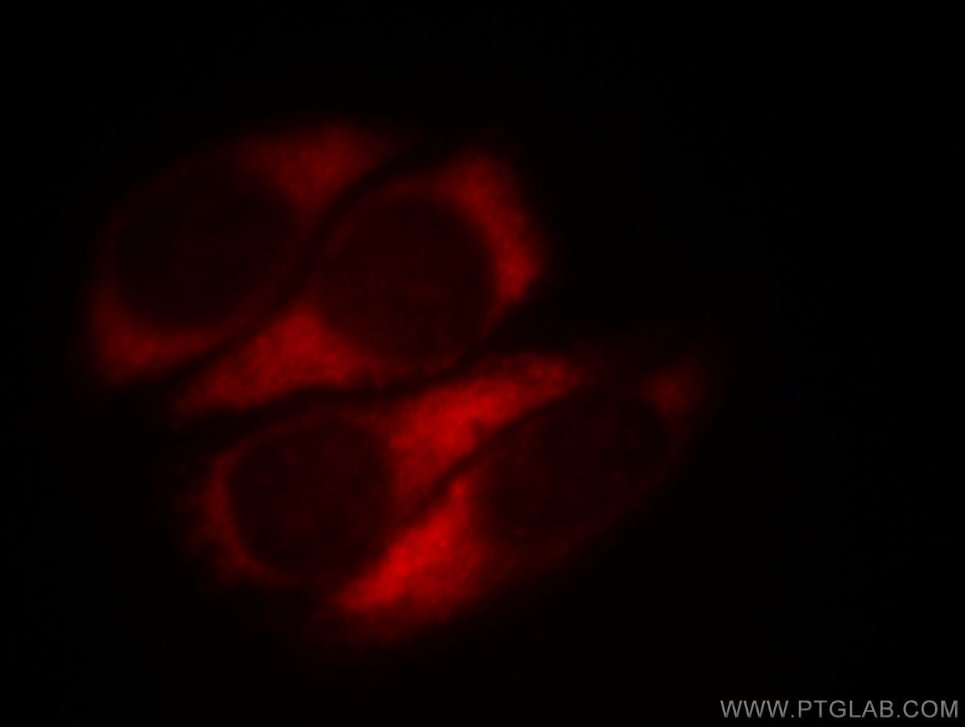
- Immunofluorescent analysis of HepG2 cells, using MTX2 antibody 11610-1-AP at 1:25 dilution and Rhodamine-labeled goat anti-rabbit IgG (red).
- Sample type
- cell line
Supportive validation
- Submitted by
- Proteintech Group (provider)
- Main image

- Experimental details
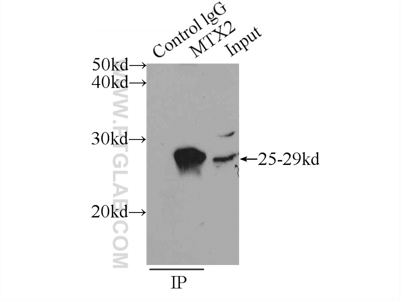
- IP Result of anti-MTX2 (IP:11610-1-AP, 3ug; Detection:11610-1-AP 1:300) with mouse heart tissue lysate 4000ug.
- Sample type
- tissue
Supportive validation
- Submitted by
- Proteintech Group (provider)
- Main image

- Experimental details
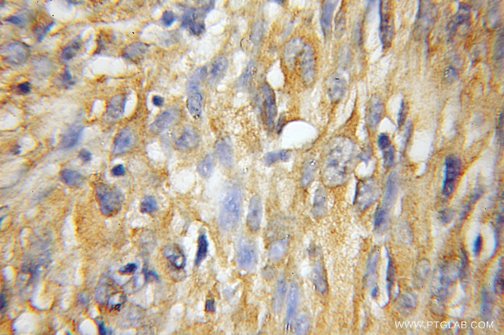
- Immunohistochemical of paraffin-embedded human gliomas using 11610-1-AP(MTX2 antibody) at dilution of 1:50 (under 10x lens)
- Sample type
- tissue