 Explore
 Validate
 Learn
Learn Immunohistochemistry
Antibody data
- Antibody Data
- Antigen structure
- References [3]
- Comments [0]
- Validations
- Immunohistochemistry [2]
- Other assay [2]
Submit
Validation data
Reference
Comment
Report error
- Product number
- MA1-22147 - Provider product page

- Provider
- Invitrogen Antibodies
- Product name
- Collagen III Monoclonal Antibody (FH-7A)
- Antibody type
- Monoclonal
- Antigen
- Other
- Description
- Recommended positive controls: human skin.
- Antibody clone number
- FH-7A
- Concentration
- 3.5 mg/mL
Submitted references Recovery of Tendon Characteristics by Inhibition of Aberrant Differentiation of Tendon-Derived Stem Cells from Degenerative Tendinopathy.
Phototherapy with low-level laser affects the remodeling of types I and III collagen in skeletal muscle repair.
Angiotensin-(1-7) ameliorates myocardial remodeling and interstitial fibrosis in spontaneous hypertension: role of MMPs/TIMPs.
Kim SJ, Oh HW, Chang JW, Kim SJ
International journal of molecular sciences 2020 Apr 13;21(8)
International journal of molecular sciences 2020 Apr 13;21(8)
Phototherapy with low-level laser affects the remodeling of types I and III collagen in skeletal muscle repair.
de Souza TO, Mesquita DA, Ferrari RA, Dos Santos Pinto D Jr, Correa L, Bussadori SK, Fernandes KP, Martins MD
Lasers in medical science 2011 Nov;26(6):803-14
Lasers in medical science 2011 Nov;26(6):803-14
Angiotensin-(1-7) ameliorates myocardial remodeling and interstitial fibrosis in spontaneous hypertension: role of MMPs/TIMPs.
Pei Z, Meng R, Li G, Yan G, Xu C, Zhuang Z, Ren J, Wu Z
Toxicology letters 2010 Nov 30;199(2):173-81
Toxicology letters 2010 Nov 30;199(2):173-81
No comments: Submit comment
Supportive validation
- Submitted by
- Invitrogen Antibodies (provider)
- Main image

- Experimental details
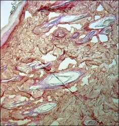
- Immunohistochemistry analysis of Collagen-III was performed in rat skin tissue using Collagen III Monoclonal Antibody (FH-7A) (Product # MA1-22147) at a dilution of 1:8,000.

- Submitted by
- Invitrogen Antibodies (provider)
- Main image
- Experimental details
- Immunohistochemistry (Paraffin) analysis of Collagen III in rat skin tissue using Collagen III Monoclonal Antibody (FH-7A) (Product # MA1-22147) at a dilution of 1:4000.

Supportive validation
- Submitted by
- Invitrogen Antibodies (provider)
- Main image
- Experimental details
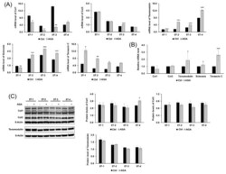
- Figure 3 Effect of AGA on tenogenic differentiation and regenerative capacity of TDSCs. ( A ) mRNA levels of collagen type I (Col1), collagen type III (Col3), tenomodulin, scleraxis, and tenascin-C were measured by qRT-PCR. ( B ) AGA increased mRNA levels of tenogenic differentiation marker in the AGA group, whereas the treatment had no effects on regenerative capacity. The relative mRNA level represents the value divided mRNA level of the AGA treatment group by mRNA level of the control group in all of the cells. ( C ) Western blot analysis was used to measure protein levels of collagen type I, collagen type III, and tenomodulin. AGA had no effects on tenogenic differentiation and regenerative capacity in the expression of protein levels. mRNA level represents mRNA expression levels standardized to beta-actin and protein level represents normalized protein expression level by beta-actin. Error bars represent the standard deviation of mean. * p < 0.05, ** p < 0.01, *** p < 0.001, compared with AGA group and control group.

- Submitted by
- Invitrogen Antibodies (provider)
- Main image

- Experimental details
- Figure 5 Effect of T0070907 on tenogenic differentiation and regenerative capacity of TDSCs. ( A ) mRNA levels of collagen type I, collagen type III, tenomodulin, scleraxis, and tenascin C were measured by qRT-PCR. ( B ) T0070907 were not increased mRNA levels of tenogenic markers. The relative mRNA level represents the value divided mRNA level of the T0070907 treatment group by mRNA level of the control group in the all of the cells. ( C ) Western blot analysis was used to measure protein levels of collagen type I, collagen type III, and tenomodulin. T0070907 had no effects on tenogenic differentiation and regenerative capacity in the expression of protein levels. mRNA level represents mRNA expression levels standardized to beta-actin and protein level represents normalized protein expression level by beta-actin. Error bars represent the standard deviation of mean. * p < 0.05, ** p < 0.01, *** p < 0.001, compared with T0070907 group and control group.
