Explore
 Validate
 Learn
Learn Western blot
 ELISA
ELISA Immunohistochemistry
Antibody data
- Antibody Data
- Antigen structure
- References [1]
- Comments [0]
- Validations
- Immunohistochemistry [1]
- Other assay [1]
Submit
Validation data
Reference
Comment
Report error
- Product number
- PA1-28870 - Provider product page

- Provider
- Invitrogen Antibodies
- Product name
- Collagen III Polyclonal Antibody
- Antibody type
- Polyclonal
- Antigen
- Other
- Description
- PA1-28870 has been successfully used in ELISA, immunohistochemistry (frozen tissue), immunoprecipitation, and Western blot applications. NOTE: The preparation of a specific antibody to Collagen III's uninterrupted "Glycine-X-Y" triplet repeat requires a non-denatured epitope format. Consequently, it is recommended to use this antibody in a Western blot using non-denaturing or "native" conditions. Store as concentrated solution. Centrifuge briefly prior to opening vial.
- Reactivity
- Human, Bovine
- Host
- Rabbit
- Isotype
- IgG
- Vial size
- 100 μg
- Concentration
- 1.16 mg/mL
- Storage
- Store at 4°C short term. For long term storage, store at -20°C, avoiding freeze/thaw cycles.
Submitted references Two-photon fluorescence and second harmonic generation characterization of extracellular matrix remodeling in post-injury murine temporomandibular joint osteoarthritis.
Reed DA, Yotsuya M, Gubareva P, Toth PT, Bertagna A
PloS one 2019;14(3):e0214072
PloS one 2019;14(3):e0214072
No comments: Submit comment
Supportive validation
- Submitted by
- Invitrogen Antibodies (provider)
- Main image
- Experimental details
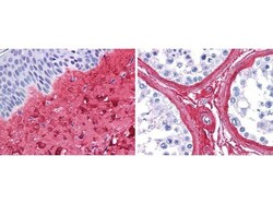
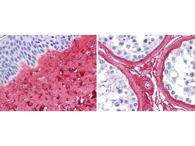
- Immunohistochemistry analysis of Collagen III was performed in human skin and testis tissue using Collagen III Polyclonal Antibody (Product # PA1-28870) at a dilution of 1:400 for 45 minutes a room temperature. The antibody showed strong staining in FFPE sections of human skin (left, dermis) with moderate to strong red staining and testis (right) where strong staining was observed within connective tissue between seminiferous tubules. The antibody showed strong extracellular staining within connective tissues across many organs with minimal background staining. Slides were steamed in 0.01 M sodium citrate buffer, pH 6.0 at 99-100°C - 20 minutes for antigen retrieval.
Supportive validation
- Submitted by
- Invitrogen Antibodies (provider)
- Main image
- Experimental details
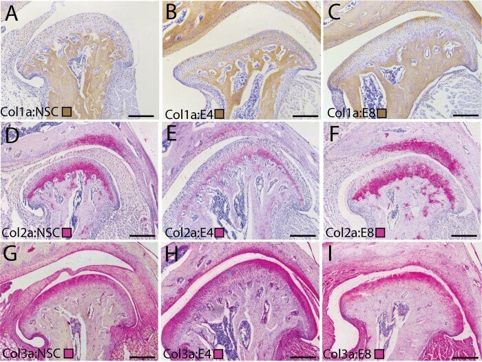
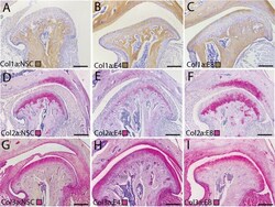
- 10.1371/journal.pone.0214072.g005 Fig 5 Changes in superficial layer collagens during early stage TMJ OA. Immunolabeling of fibrillar collagens in the TMJ illustrating changes in the composition of the superficial layer extracellular matrix that accompanies the progression of early stage TMJ-OA. Collagen I (A-C) is immunolabeled using a brown chromogen. Collagen II (D-F) and Collagen III (G-I) are immunolabeled using a pink chromogen. Cell nuclei are labeled in purple with hematoxylin. Tissues from non-surgical controls (A, D, G) are compared with 4 (B, E, H) and 8 (C, F, I) week post-injury tissues. All images are representative of their experimental group (n = 3) and taken from homologous regions of the mandibular condyle. Differences in the shape of the condylar reflect variable amounts of condylar flattening, typical of the surgical model. Medial is to the right in all joints. Scale bars equal 50 mum. Note strong staining for Collagens I/III in the superficial layer in post-injury tissues, with Collagen II restricted to the deeper chondroblastic/hypertrophic layers. Primary and secondary antibody controls were negative.