 Explore
 Validate
 Learn
Learn Western blot
Antibody data
- Antibody Data
- Antigen structure
- References [4]
- Comments [0]
- Validations
- Western blot [8]
- Immunohistochemistry [1]
Submit
Validation data
Reference
Comment
Report error
- Product number
- GTX102997 - Provider product page

- Provider
- GeneTex
- Proper citation
- GeneTex Cat#GTX102997, RRID:AB_10618014
- Product name
- Collagen III antibody [C2C3], C-term
- Antibody type
- Polyclonal
- Reactivity
- Human, Mouse, Rat
- Host
- Rabbit
Submitted references Autologous Adipose-Derived Tissue Matrix Part I: Biologic Characteristics.
The Implication of Substance P in the Development of Tendinopathy: A Case Control Study.
Simulated physiological stretch increases expression of extracellular matrix proteins in human bladder smooth muscle cells via integrin α4/αv-FAK-ERK1/2 signaling pathway.
(-)-Epicatechin improves mitochondrial-related protein levels and ameliorates oxidative stress in dystrophic δ-sarcoglycan null mouse striated muscle.
Schendel SA
Aesthetic surgery journal 2017 Oct 1;37(9):1062-1068
Aesthetic surgery journal 2017 Oct 1;37(9):1062-1068
The Implication of Substance P in the Development of Tendinopathy: A Case Control Study.
Han SH, Choi W, Song J, Kim J, Lee S, Choi Y, Byun SE, Ahn T, Ahn H, Ding C, Baik L, Ward S, Ting K, Lee S
International journal of molecular sciences 2017 Jun 9;18(6)
International journal of molecular sciences 2017 Jun 9;18(6)
Simulated physiological stretch increases expression of extracellular matrix proteins in human bladder smooth muscle cells via integrin α4/αv-FAK-ERK1/2 signaling pathway.
Chen S, Peng C, Wei X, Luo D, Lin Y, Yang T, Jin X, Gong L, Li H, Wang K
World journal of urology 2017 Aug;35(8):1247-1254
World journal of urology 2017 Aug;35(8):1247-1254
(-)-Epicatechin improves mitochondrial-related protein levels and ameliorates oxidative stress in dystrophic δ-sarcoglycan null mouse striated muscle.
Ramirez-Sanchez I, De los Santos S, Gonzalez-Basurto S, Canto P, Mendoza-Lorenzo P, Palma-Flores C, Ceballos-Reyes G, Villarreal F, Zentella-Dehesa A, Coral-Vazquez R
The FEBS journal 2014 Dec;281(24):5567-80
The FEBS journal 2014 Dec;281(24):5567-80
No comments: Submit comment
Supportive validation
- Submitted by
- GeneTex (provider)
- Main image

- Experimental details
- Collagen III antibody [C2C3], C-term detects Collagen III protein by western blot analysis.A. 30 ?g Neuro2A whole lysate/extract B. 30 ?g NIH-3T3 whole cell lysate/extract C. 30 ?g BCL-1 whole cell lysate/extract D. 30 ?g Raw264.7 whole cell lysate/extract E. 30 ?g C2C12 whole cell lysate/extract5% SDS-PAGECollagen III antibody [C2C3], C-term (GTX102997) dilution: 1:1000 The HRP-conjugated anti-rabbit IgG antibody (GTX213110-01) was used to detect the primary antibody.

- Submitted by
- GeneTex (provider)
- Main image

- Experimental details
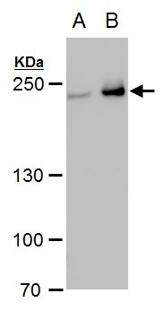
- Collagen III antibody [C2C3], C-term detects Collagen III protein by western blot analysis.A. 50 ?g rat liver lysate/extract 5% SDS-PAGECOL3A1 antibody [C2C3], C-term (GTX102997) dilution: 1:1000 The HRP-conjugated anti-rabbit IgG antibody (GTX213110-01) was used to detect the primary antibody.

- Submitted by
- GeneTex (provider)
- Main image

- Experimental details
- Sample (30 ug of whole cell lysate) A: Hela 5% SDS PAGE GTX102997 diluted at 1:1000
- Validation comment
- WB

- Submitted by
- GeneTex (provider)
- Main image

- Experimental details
- Collagen III antibody [C2C3], C-term detects Collagen III protein by western blot analysis.A. 30 ?g PC-12 whole cell extract B. 30 ?g Rat2 whole cell extract5% SDS-PAGECollagen III antibody [C2C3], C-term (GTX102997) dilution: 1:1000 The HRP-conjugated anti-rabbit IgG antibody (GTX213110-01) was used to detect the primary antibody.
- Submitted by
- GeneTex (provider)
- Main image

- Experimental details
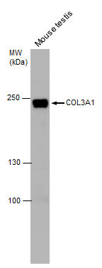
- Collagen III antibody [C2C3], C-term detects Collagen III protein by western blot analysis. Mouse tissue extracts (30 ?g) was separated by 5% SDS-PAGE, and the membrane was blotted with Collagen III antibody [C2C3], C-term (GTX102997) at a dilution of 1:1000. The HRP-conjugated anti-rabbit IgG antibody (GTX213110-01) was used to detect the primary antibody.
- Submitted by
- GeneTex (provider)
- Main image

- Experimental details
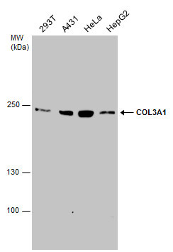
- COL3A1 antibody [C2C3], C-term detects COL3A1 protein by western blot analysis. Various whole cell extracts (30 ?g) were separated by 5% SDS-PAGE, and the membrane was blotted with COL3A1 antibody [C2C3], C-term (GTX102997) diluted at a dilution of 1:1000.
- Validation comment
- WB

- Submitted by
- GeneTex (provider)
- Main image

- Experimental details
- Various whole cell extracts (30 ?g) were separated by 5% SDS-PAGE, and the membrane was blotted with COL3A1 antibody [C2C3], C-term (GTX102997) diluted at 1:1000. The HRP-conjugated anti-rabbit IgG antibody (GTX213110-01) was used to detect the primary antibody.
- Submitted by
- GeneTex (provider)
- Main image

- Experimental details
- Various whole cell extracts (30 ?g) were separated by 5% SDS-PAGE, and the membrane was blotted with Collagen III antibody [C2C3], C-term (GTX102997) diluted at 1:5000. The HRP-conjugated anti-rabbit IgG antibody (GTX213110-01) was used to detect the primary antibody.

Supportive validation
- Submitted by
- GeneTex (provider)
- Main image
- Experimental details
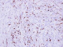
- Immunohistochemical analysis of paraffin-embedded human PAPILLARY CA_STROMAL CELLS, using Collagen III (GTX102997) antibody at 1:250 dilution.
