Explore
 Validate
 Learn
Learn Western blot
 ELISA
ELISAAntibody data
- Antibody Data
- Antigen structure
- References [6]
- Comments [0]
- Validations
- Western blot [1]
- Immunohistochemistry [1]
Submit
Validation data
Reference
Comment
Report error
- Product number
- GTX27778 - Provider product page

- Provider
- GeneTex
- Proper citation
- GeneTex Cat#GTX27778, RRID:AB_374551
- Product name
- Collagen III antibody
- Antibody type
- Polyclonal
- Reactivity
- Human, Bovine
- Host
- Rabbit
Submitted references KCTD11 inhibits growth and metastasis of hepatocellular carcinoma through activating Hippo signaling.
Saikosaponin d induces cell death through caspase-3-dependent, caspase-3-independent and mitochondrial pathways in mammalian hepatic stellate cells.
Candesartan ameliorates arsenic-induced hypertensive vascular remodeling by regularizing angiotensin II and TGF-beta signaling in rats.
Vasoprotective effects of urocortin 1 against atherosclerosis in vitro and in vivo.
The behavior of ligament cells cultured on elastin and collagen scaffolds.
Impact of Smad3 loss of function on scarring and adhesion formation during tendon healing.
Tong R, Yang B, Xiao H, Peng C, Hu W, Weng X, Cheng S, Du C, Lv Z, Ding C, Zhou L, Xie H, Wu J, Zheng S
Oncotarget 2017 Jun 6;8(23):37717-37729
Oncotarget 2017 Jun 6;8(23):37717-37729
Saikosaponin d induces cell death through caspase-3-dependent, caspase-3-independent and mitochondrial pathways in mammalian hepatic stellate cells.
Chen MF, Huang SJ, Huang CC, Liu PS, Lin KI, Liu CW, Hsieh WC, Shiu LY, Chen CH
BMC cancer 2016 Jul 26;16:532
BMC cancer 2016 Jul 26;16:532
Candesartan ameliorates arsenic-induced hypertensive vascular remodeling by regularizing angiotensin II and TGF-beta signaling in rats.
Khuman MW, Harikumar SK, Sadam A, Kesavan M, Susanth VS, Parida S, Singh KP, Sarkar SN
Toxicology 2016 Dec 30;374:29-41
Toxicology 2016 Dec 30;374:29-41
Vasoprotective effects of urocortin 1 against atherosclerosis in vitro and in vivo.
Hasegawa A, Sato K, Shirai R, Watanabe R, Yamamoto K, Watanabe K, Nohtomi K, Hirano T, Watanabe T
PloS one 2014;9(12):e110866
PloS one 2014;9(12):e110866
The behavior of ligament cells cultured on elastin and collagen scaffolds.
Mizutani N, Kageyama S, Yamada M, Hasegawa M, Miyamoto K, Horiuchi T
Journal of artificial organs : the official journal of the Japanese Society for Artificial Organs 2014 Mar;17(1):50-9
Journal of artificial organs : the official journal of the Japanese Society for Artificial Organs 2014 Mar;17(1):50-9
Impact of Smad3 loss of function on scarring and adhesion formation during tendon healing.
Katzel EB, Wolenski M, Loiselle AE, Basile P, Flick LM, Langstein HN, Hilton MJ, Awad HA, Hammert WC, O'Keefe RJ
Journal of orthopaedic research : official publication of the Orthopaedic Research Society 2011 May;29(5):684-93
Journal of orthopaedic research : official publication of the Orthopaedic Research Society 2011 May;29(5):684-93
No comments: Submit comment
Supportive validation
- Submitted by
- GeneTex (provider)
- Main image

- Experimental details
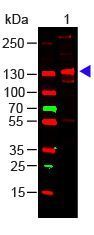
- Western Blot of Rabbit Anti-Collagen III Antibody (GTX27778) Load: 100 ng Human Collagen III. Primary antibody: Collagen III Antibody at 1:1000 o/n at 4¢XC. Secondary antibody: DyLight? 649 Goat anti-rabbit at 1:20,000 for 30 min at RT Block for 30 min at RT. Predicted/Observed size: 138 kDa, 138 kDa.
Supportive validation
- Submitted by
- GeneTex (provider)
- Main image
- Experimental details
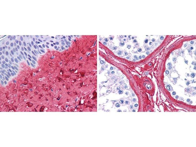
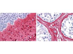
- GeneTex anti collagen III antibody (GTX27778, 45 min RT) showed strong staining in FFPE sections of human skin(left, dermis) with moderate to strong red staining and testis (right) where strong staining was observed within connective tissue between seminiferous tubules. The antibody showed strong extracellular staining within connective tissues across many organs with minimal background staining. Slides were steamed in 0.01 M sodium citrate buffer, pH 6.0 at 99-100¢XC - 20 minutes for antigen retrieval.