Explore
 Validate
 Learn
Learn Western blot
 ELISA
ELISAAntibody data
- Antibody Data
- Antigen structure
- References [1]
- Comments [0]
- Validations
- Western blot [1]
- Immunohistochemistry [2]
Submit
Validation data
Reference
Comment
Report error
- Product number
- R1040X - Provider product page

- Provider
- OriGene
- Product name
- Collagen III (COL3A1) rabbit polyclonal antibody, Aff - Purified
- Antibody type
- Polyclonal
- Description
- Collagen III (COL3A1) rabbit polyclonal antibody, Aff - Purified
- Host
- Rabbit
- Conjugate
- Unconjugated
- Epitope
- COL3A1
- Isotype
- IgG
- Antibody clone number
- NULL
- Vial size
- 500 µg
- Concentration
- 1.0 mg/ml (by UV absorbance at 280 nm)
Submitted references Umbilical cord as human cell source for mitral valve tissue engineering - venous vs. arterial cells.
Malischewski A, Moreira R, Hurtado L, Gesché V, Schmitz-Rode T, Jockenhoevel S, Mela P
Biomedizinische Technik. Biomedical engineering 2017 Oct 26;62(5):457-466
Biomedizinische Technik. Biomedical engineering 2017 Oct 26;62(5):457-466
No comments: Submit comment
Supportive validation
- Submitted by
- OriGene (provider)
- Main image

- Experimental details
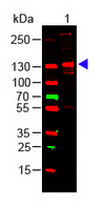
- Western blot of rabbit collagen III alpha 1 chain antibody .?Lane 1: human collagen III load: 100 ng per lane, primary antibody: Collagen III antibody at 1:1000 o/n at 4°C, secondary antibody: DyLight?TM 649 goat anti-rabbit.
- Validation comment
- WB
Supportive validation
- Submitted by
- OriGene (provider)
- Main image

- Experimental details
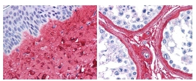
- R1040 Collagen III Antibody (1/400, 45 min RT) showed strong staining in FFPE sections of Human skin (Left, dermis) with moderate to strong Red staining and testis (Right) where strong staining was observed within connective tissue between seminiferous tubules. The antibody showed strong extracellular staining within connective tissues across many organs with minimal background staining. Slides were steamed in 0.01 M sodium citrate buffer, pH 6.0 at 99-100°C - 20 minutes for antigen retrieval. Images provided courtesy of LifeSpan Biosciences, Seattle, WA
- Validation comment
- IHC
- Submitted by
- OriGene (provider)
- Main image
- Experimental details
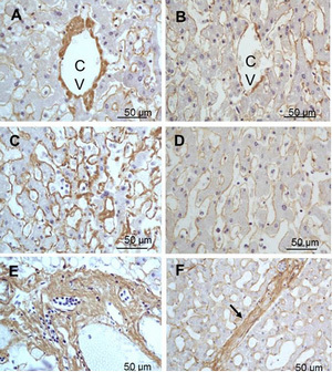
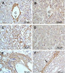
- Immunohistochemistry of tissue of right lobe of the liver section (Formalin-fixed, Paraffin-embedded). A:Central Vein (CV) fibrosis, B: Non-fibrotic CV, C: Perisinusodial fibrosis, D: Non-fibrotic area, E: Protat tract fibrosis, F: Septal fibrosis (arrow)?using Collagen type III alpha 1 chain antibody .?R1040 at 1:500 for 4°C for 24hr. Secondary antibody: Peroxidase biotin-streptavidin rabbit secondary antibody and DAB as chromogen. Nuclei were counterstained purple with hematoxylin.
- Validation comment
- IHC