 Explore
 Validate
 Learn
Learn Western blot
 ELISA
ELISAAntibody data
- Antibody Data
- Antigen structure
- References [0]
- Comments [0]
- Validations
- Western blot [1]
- Immunohistochemistry [2]
Submit
Validation data
Reference
Comment
Report error
- Product number
- LS-C745277 - Provider product page

- Provider
- LSBio
- Product name
- COL3A1 / Collagen III Antibody LS-C745277
- Antibody type
- Polyclonal
- Reactivity
- Human, Bovine
- Host
- Rabbit
- Isotype
- IgG
- Storage
- Store vial at -20°C or below prior to opening. Dilute 1:10 to minimize loss. Store the vial at -20°C or below after dilution. Avoid freeze-thaw cycles.
No comments: Submit comment
Supportive validation
- Submitted by
- LSBio (provider)
- Main image
- Experimental details
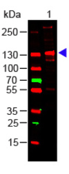
- Western Blot of rabbit Anti-COLLAGEN III Antibody Lane 1: Human Collagen III Load: 100 ng per lane Primary antibody: Collagen III Antibody at 1:1000 o/n at 4°C Secondary antibody: DyLight 649 Goat anti-rabbit at 1:20,000 for 30 min at RT Block: MB-070 for 30 min at RT Predicted/Observed size: 138 kDa, 138 kDa

Supportive validation
- Submitted by
- LSBio (provider)
- Main image
- Experimental details
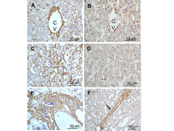
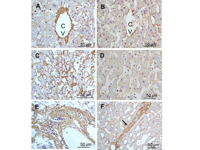
- Immunohistochemistry of rabbit Anti-collagen type III antibody. Tissue: right lobe of the liver section. A:Central Vein (CV) fibrosis, B: Non-fibrotic CV, C: Perisinusodial fibrosis, D: Non-fibrotic area, E: Protat tract fibrosis, F: Septal fibrosis (arrow). Fixation: formalin fixed paraffin embedded. Antigen retrieval: not required. Primary antibody: Anti-collagen type III at 1:500 for 4°C for 24hr. Secondary antibody: Peroxidase biotin-streptavidin rabbit secondary antibody at 1:10,000 for 45 min at RT. Localization: Anti-collagen type III is intra and extracellular. Staining: 3.3’-diaminobenzidine tetrahydrochloride was used as the chromogen. Nuclei were counterstained purple with hematoxylin.
- Submitted by
- LSBio (provider)
- Main image

- Experimental details
- Anti collagen III antibody showed strong staining in FFPE sections of human skin(left, dermis) with moderate to strong red staining and testis (right) where strong staining was observed within connective tissue between seminiferous tubules. The antibody showed strong extracellular staining within connective tissues across many organs with minimal background staining. Slides were steamed in 0.01 M sodium citrate buffer, pH 6.0 at 99-100°C - 20 minutes for antigen retrieval.
