Explore
 Validate
 Learn
Learn Western blot
 ELISA
ELISA Immunohistochemistry
Antibody data
- Antibody Data
- Antigen structure
- References [1]
- Comments [0]
- Validations
- Immunohistochemistry [2]
- Other assay [2]
Submit
Validation data
Reference
Comment
Report error
- Product number
- MA5-15594 - Provider product page

- Provider
- Invitrogen Antibodies
- Product name
- BDH1 Monoclonal Antibody (1A5)
- Antibody type
- Monoclonal
- Antigen
- Purifed from natural sources
- Description
- MA5-15594 targets BDH1 in indirect ELISA, IHC and WB applications and shows reactivity with Human and mouse samples. The MA5-15594 immunogen is purified recombinant fragment of human BDH1 expressed in E. Coli. MA5-15594 detects BDH1 which has a predicted molecular weight of approximately 38kDa.
- Reactivity
- Human, Mouse
- Host
- Mouse
- Isotype
- IgG
- Antibody clone number
- 1A5
- Vial size
- 100 μL
- Concentration
- Conc. Not Determined
- Storage
- Store at 4°C short term. For long term storage, store at -20°C, avoiding freeze/thaw cycles.
Submitted references E2F6 Impairs Glycolysis and Activates BDH1 Expression Prior to Dilated Cardiomyopathy.
Major JL, Dewan A, Salih M, Leddy JJ, Tuana BS
PloS one 2017;12(1):e0170066
PloS one 2017;12(1):e0170066
No comments: Submit comment
Supportive validation
- Submitted by
- Invitrogen Antibodies (provider)
- Main image

- Experimental details
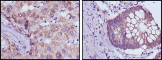
- Immunohistochemical analysis of paraffin-embedded human liver cancer (left) and colorectal cancer tissues (right) using BDH1 monoclonal antibody (Product # MA5-15594) followed with DAB staining.
- Submitted by
- Invitrogen Antibodies (provider)
- Main image
- Experimental details
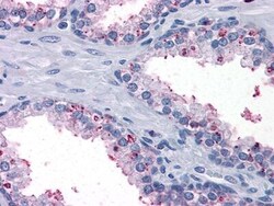
- Immunohistochemical analysis of paraffin-embedded human prostate tissues using BDH1 monoclonal antibody (Product # MA5-15594).

Supportive validation
- Submitted by
- Invitrogen Antibodies (provider)
- Main image

- Experimental details
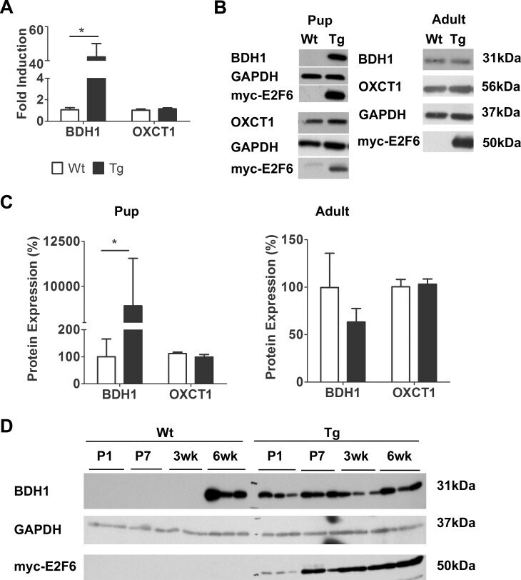
- Fig 1 E2F6 Activates BDH1 Expression in Neonatal Myocardium. ( A) Bdh1 (beta-hydroxybutyrate dehydrogenase 1) and Oxct1 (succinyl-CoA: 3-oxoacid-CoA transferase) transcript levels in Wt and Tg myocardium 7 days after birth. Expression is normalized to Gapdh . Results represent mean+-SEM values (n = 5-7). ( B) Representative immunoblots of protein from Wt and Tg mouse myocardium at post-natal day 1 (pups) and 6wks of age (adult) examined with the anti-BDH1 and anti-OXCT1. ( C) Densitometric quantification of BDH1 and OXCT1 expression from immunoblots. Expression is normalized against GAPDH. Results represent mean+-SEM values (n = 4). ( D) Immunoblot of protein from Wt and Tg myocardium with BDH1 at the indicated time points after birth.* P
- Submitted by
- Invitrogen Antibodies (provider)
- Main image
- Experimental details
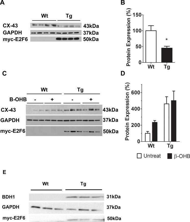
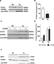
- Fig 5 beta-OHB Regulates CX-43 Protein Expression in Neonatal Cardiomyocytes. ( A) Representative immunoblot of protein isolated from Wt and Tg myocardium at post-natal day 1 with anti-CX-43. ( B) Densitometric quantification of CX-43 immunoblot. Expression is normalized against GAPDH. Results represent mean+-SEM values (n = 4). ( C) Representative immunoblot analyses of protein from Wt and Tg neonatal cardiomyocytes following incubation with or without beta-OHB (beta-hydroxybutyrate). ( D) Densitometric quantification of CX-43 in treated and non-treated neonatal cardiomyocytes. Expression is normalized against GAPDH. Results represent the mean+-SEM values (n = 3-5). A two-way ANOVA was used to measure statistical significance. ( E ) Representative immunoblot analysis of BDH1 in Wt and Tg neonatal cardiomyocytes. * P