Explore
 Validate
 Learn
Learn Western blot
 Immunohistochemistry
Antibody data
- Antibody Data
- Antigen structure
- References [0]
- Comments [0]
- Validations
- Western blot [1]
Submit
Validation data
Reference
Comment
Report error
- Product number
- BS2011 - Provider product page

- Provider
- Bioworld Technology, Inc
- Proper citation
- Bioworld Technology Cat#BS2011, RRID:AB_1662917
- Product name
- MNAT1 (E123) polyclonal antibody
- Antibody type
- Polyclonal
- Antigen
- Synthetic peptide, corresponding to amino acids 100-150 of Human MNAT1.
- Description
- Progression through the cell cycle requires activation of a series of enzymes designated cyclin dependent kinases (Cdks). The monomeric catalytic subunit, Cdk2, a critical enzyme for initiation of cell cycle progression, is completely inactive. Partial activation is achieved by the binding of regulatory cyclins such as cyclin D1, while full activation requires phosphorylation at Thr 160. The enzyme responsible for phosphorylation of Thr 160 in Cdk2 and also Thr 161 in Cdc2 p34, designated Cdk-activating kinase (CAK), has been partially purified and shown to be comprised of a catalytic subunit, a regulatory subunit and a subunit of unknown function. The regulatory subunit is a novel cyclin (cyclin H) and is required for activation of Cdk7. This previously undescribed protein, now termed Mat1 p36, has been cloned as a protein that associates with the cyclin H/Cdk7 nuclear complex at all stages of the cell cycle. Cyclin H/Cdk7/Mat1 p36 complexes display kinase activity towards Cdk activation domains, and the carboxy terminus of RNA polymerase II. Mat1 p36 appears to constitute the first example of an assembly factor, essential for the formation of an active Cdk/cyclin complex.
- Reactivity
- Human, Mouse, Rat
- Host
- Rabbit
- Isotype
- IgG
- Vial size
- 100ul
- Concentration
- 1 mg/ml
- Storage
- Store at 4°C short term. Aliquot and store at -20°C long term. Avoid freeze-thaw cycles.
No comments: Submit comment
Supportive validation
- Submitted by
- Bioworld Technology, Inc (provider)
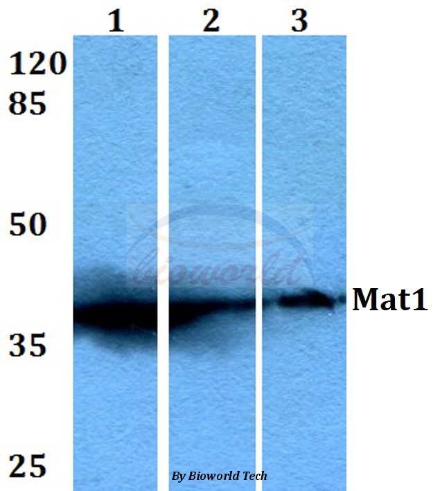
- Main image

- Experimental details
- Western blot (WB) analysis of Mat1 (E123) pAb at 1:500 dilutionLane1:HEK293T whole cell lysateLane2:Raw264.7 whole cell lysateLane3:PC12 whole cell lysate