Explore
 Validate
 Learn
Learn Western blot
Antibody data
- Antibody Data
- Antigen structure
- References [1]
- Comments [0]
- Validations
- Western blot [4]
- Immunocytochemistry [1]
- Immunohistochemistry [1]
Submit
Validation data
Reference
Comment
Report error
- Product number
- GTX113312 - Provider product page

- Provider
- GeneTex
- Proper citation
- GeneTex Cat#GTX113312, RRID:AB_2036200
- Product name
- AKT3 antibody
- Antibody type
- Polyclonal
- Reactivity
- Human, Mouse
- Host
- Rabbit
Submitted references Erythropoietin protects neuroblastoma cells against etoposide and vincristine by activating ERK and AKT pathways but has no effect in kidney cells.
Vazquez-Mellado MJ, Aguilar C, Rocha-Zavaleta L
Life sciences 2015 Sep 15;137:142-9
Life sciences 2015 Sep 15;137:142-9
No comments: Submit comment
Supportive validation
- Submitted by
- GeneTex (provider)
- Main image

- Experimental details
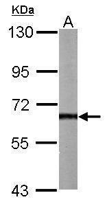
- Sample (50 ?g of whole cell lysate) A: Mouse brain 7.5% SDS PAGE GTX113312 diluted at 1:1000 The HRP-conjugated anti-rabbit IgG antibody (GTX213110-01) was used to detect the primary antibody.
- Submitted by
- GeneTex (provider)
- Main image
- Experimental details
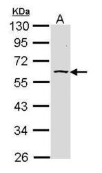
- Sample (30 ?g of whole cell lysate) A: Raji 10% SDS PAGE GTX113312 diluted at 1:1000 The HRP-conjugated anti-rabbit IgG antibody (GTX213110-01) was used to detect the primary antibody.
- Submitted by
- GeneTex (provider)
- Main image

- Experimental details
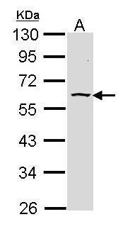
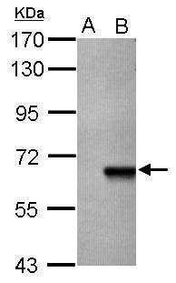
- Sample (20 ug of whole cell lysate) A: HeLa cell lysate B: HeLa cell transfect with AKT 7.5% SDS PAGE GTX113312 diluted at 1:5000
- Validation comment
- WB
- Submitted by
- GeneTex (provider)
- Main image
- Experimental details
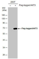
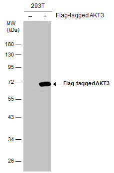
- Non-transfected (¡V) and transfected (+) 293T whole cell extracts (30 ?g) were separated by 10% SDS-PAGE, and the membrane was blotted with AKT3 antibody (GTX113312) diluted at 1:1000. The HRP-conjugated anti-rabbit IgG antibody (GTX213110-01) was used to detect the primary antibody.
Supportive validation
- Submitted by
- GeneTex (provider)
- Main image

- Experimental details
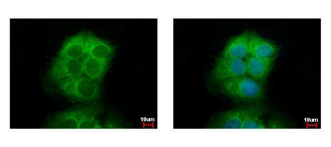
- Akt3 antibody detects Akt3 protein at cytoplasm by immunofluorescent analysis. Sample: MCF-7 cells were fixed in ice-cold MeOH for 5 min.Green: Akt3 protein stained by Akt3 antibody (GTX113312) diluted at 1:500.Blue: Hoechst 33342 staining.
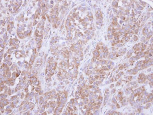
Supportive validation
- Submitted by
- GeneTex (provider)
- Main image
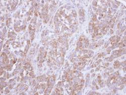
- Experimental details
- Immunohistochemical analysis of paraffin-embedded A549 xenograft, using Akt3(GTX113312) antibody at 1:500 dilution.