Explore
 Validate
 Learn
Learn Western blot
 ELISA
ELISAAntibody data
- Antibody Data
- Antigen structure
- References [0]
- Comments [0]
- Validations
- Western blot [4]
- Immunocytochemistry [2]
- Immunohistochemistry [4]
- Flow cytometry [1]
Submit
Validation data
Reference
Comment
Report error
- Product number
- R33341 - Provider product page

- Provider
- NSJ Bioreagents
- Product name
- AKT3 Antibody
- Antibody type
- Polyclonal
- Description
- This highly specific AKT3 antibody is suitable for use in Western blot/Immunohistochemistry/Flow cytometry/Immunofluorescence/ELISA applications with human, mouse and rat samples.
- Reactivity
- Human, Mouse, Rat
- Host
- Goat
- Conjugate
- Unconjugated
- Vial size
- 100 ug
- Concentration
- 0.5 mg/ml in 1X TBS, pH7.3, with 0.5% BSA (US sourced) and 0.02% sodium azide
- Storage
- Aliquot and store the AKT3 antibody at -20oC.
No comments: Submit comment
Supportive validation
- Submitted by
- NSJ Bioreagents (provider)
- Main image
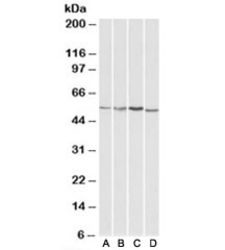
- Experimental details
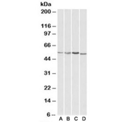
- Western blot testing of HepG2 [A], Jurkat [B], mouse brain [C] and rat brain [D] lysates with AKT3 antibody at 1ug/ml. Predicted molecular weight: ~56kDa but can be observed from 60~65kDa.
- Submitted by
- NSJ Bioreagents (provider)
- Main image

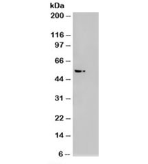
- Experimental details
- Western blot of HEK293 lysate overexpressing AKT3 probed with AKT3 antibody (mock transfection on right). Predicted molecular weight: ~56kDa but can be observed from 60~65kDa.
- Submitted by
- NSJ Bioreagents (provider)
- Main image
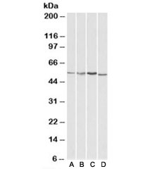
- Experimental details
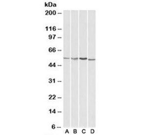
- Western blot testing of HepG2 [A], Jurkat [B], mouse brain [C] and rat brain [D] lysates with AKT3 antibody at 1ug/ml. Predicted molecular weight: ~56kDa but can be observed from 60~65kDa.
- Submitted by
- NSJ Bioreagents (provider)
- Main image
- Experimental details
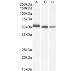
- Western blot of 1A) human thyroid, 2) mouse brain and 3) rat brain tissue lysate probed with AKT3 antibody. Predicted molecular weight: ~56 kDa but can be observed from 60~65 kDa.

Supportive validation
- Submitted by
- NSJ Bioreagents (provider)
- Main image
- Experimental details
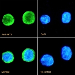
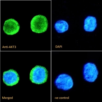
- IF/ICC staining of fixed and permeabilized human ThP-1 cells with AKT3 antibody (green) at 10ug/ml and DAPI nuclear stain (blue).
- Submitted by
- NSJ Bioreagents (provider)
- Main image
- Experimental details
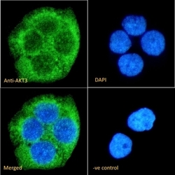
- IF/ICC staining of fixed and permeabilized human A431 cells with AKT3 antibody (green) at 10ug/ml and DAPI nuclear stain (blue).

Supportive validation
- Submitted by
- NSJ Bioreagents (provider)
- Main image

- Experimental details
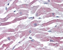
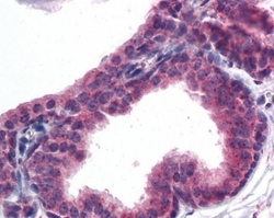
- IHC testing of FFPE human prostate tissue with AKT3 antibody at 5ug/ml. Required HIER: steamed antigen retrieval with pH6 citrate buffer; AP-staining.

- Submitted by
- NSJ Bioreagents (provider)
- Main image
- Experimental details
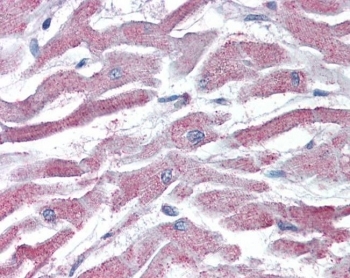
- IHC testing of FFPE human heart tissue with AKT3 antibody at 5ug/ml. Required HIER: steamed antigen retrieval with pH6 citrate buffer; AP-staining.
- Submitted by
- NSJ Bioreagents (provider)
- Main image
- Experimental details
- IHC testing of FFPE human prostate tissue with AKT3 antibody at 5ug/ml. Required HIER: steamed antigen retrieval with pH6 citrate buffer; AP-staining.

- Submitted by
- NSJ Bioreagents (provider)
- Main image

- Experimental details
- IHC testing of FFPE human heart tissue with AKT3 antibody at 5ug/ml. Required HIER: steamed antigen retrieval with pH6 citrate buffer; AP-staining.

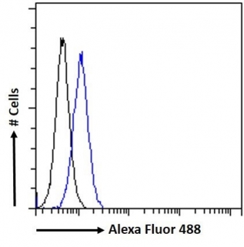
Supportive validation
- Submitted by
- NSJ Bioreagents (provider)
- Main image
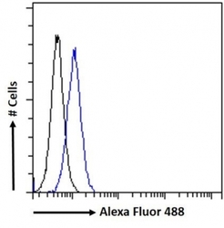
- Experimental details
- FACS testing of fixed and permeabilized human HepG2 cells with AKT3 antibody (blue) at 10ug/ml and naive goat Ig (black).