Explore
 Validate
 Learn
Learn Western blot
Antibody data
- Antibody Data
- Antigen structure
- References [0]
- Comments [0]
- Validations
- Western blot [1]
- Immunocytochemistry [2]
- Immunohistochemistry [3]
- Flow cytometry [1]
Submit
Validation data
Reference
Comment
Report error
- Product number
- PA5-109345 - Provider product page

- Provider
- Invitrogen Antibodies
- Product name
- AKT3 Polyclonal Antibody
- Antibody type
- Polyclonal
- Antigen
- Recombinant full-length protein
- Description
- Positive Control: SH-SY5Y cell lysates, A549, EA.hy926, human thyroid tissue, human kidney tissue, mouse brain tissue.
- Reactivity
- Human
- Host
- Rabbit
- Isotype
- IgG
- Vial size
- 100 µL
- Concentration
- 1 mg/mL
- Storage
- -20° C, Avoid Freeze/Thaw Cycles, store in dark
No comments: Submit comment
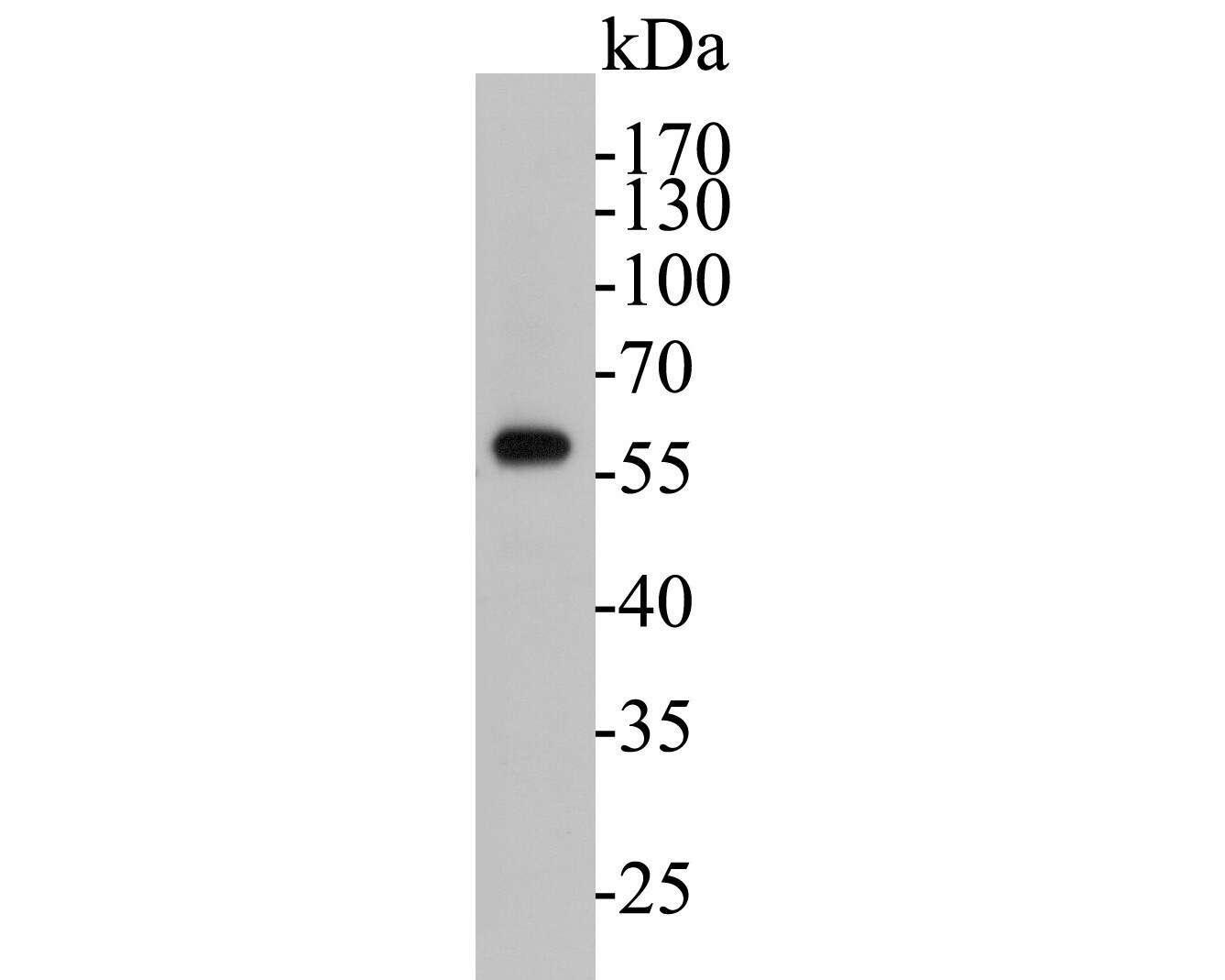
Supportive validation
- Submitted by
- Invitrogen Antibodies (provider)
- Main image

- Experimental details
- Western blot analysis of AKT3 in SH-SY5Y cell lysate. Samples were transferred to PVDF membrane, blocked with 5% BSA (1 hour), incubated with AKT3 polyclonal antibody (Product # PA5-109345), at a dilution of 1:500, followed by Goat Anti-Rabbit IgG-HRP (1 hour) with a dilution of 1:5000.
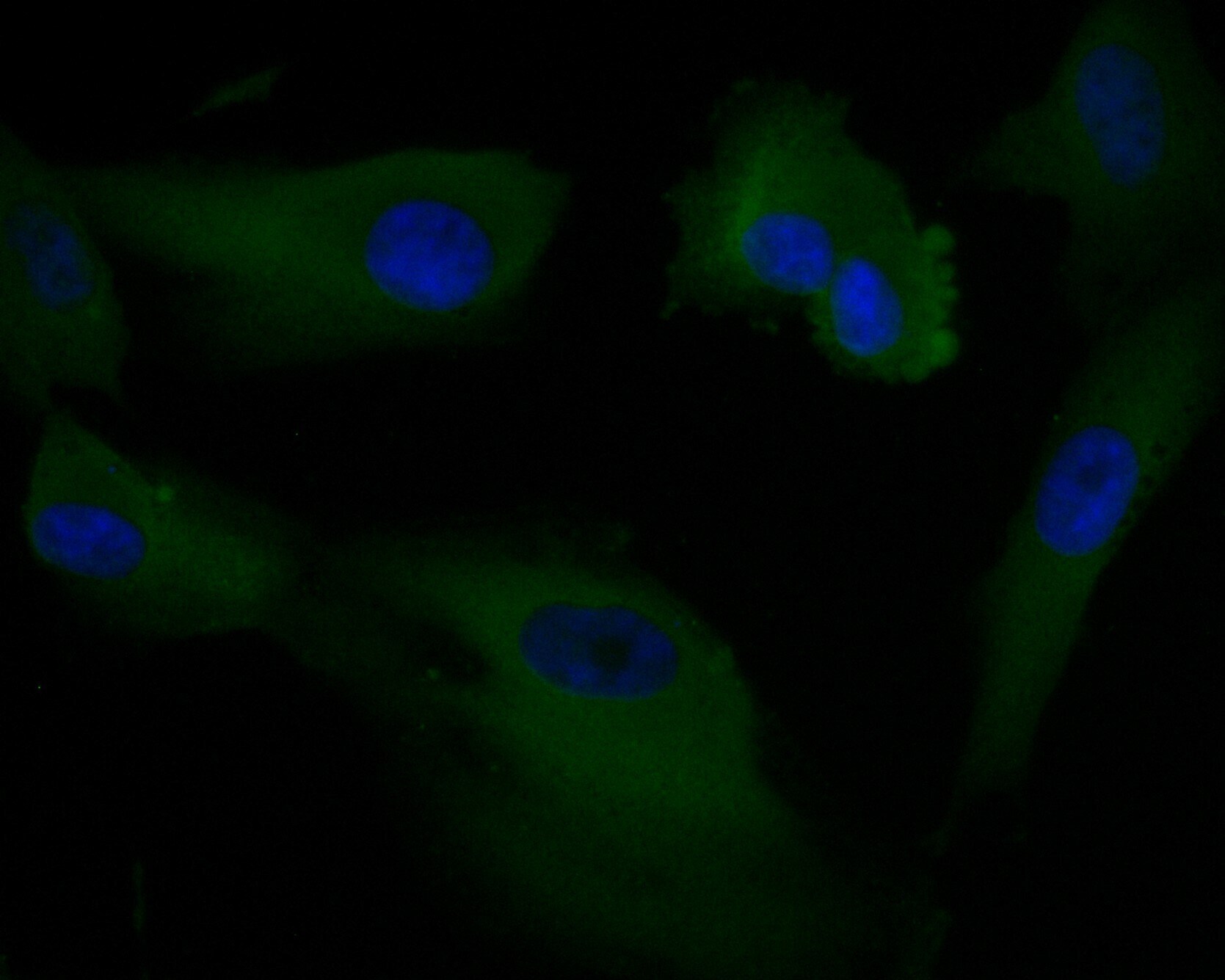
Supportive validation
- Submitted by
- Invitrogen Antibodies (provider)
- Main image
- Experimental details
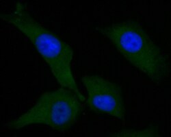
- Immunofluorescent analysis of AKT3 in EA.hy926 cells (green). Samples were formalin fixed, permeabilized with 0.1% Triton X-100 in TBS (1 hour, room temperature) and blocked with 1% BSA (15 min, room temperature), incubated with AKT3 polyclonal antibody (Product # PA5-109345) at a dilution of 1:50 (1 hour, room temperature), and followed by Alexa Fluor 488 Goat anti-Rabbit IgG and DAPI (blue) with a dilution of 1:100.
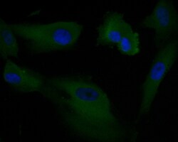
- Submitted by
- Invitrogen Antibodies (provider)
- Main image
- Experimental details
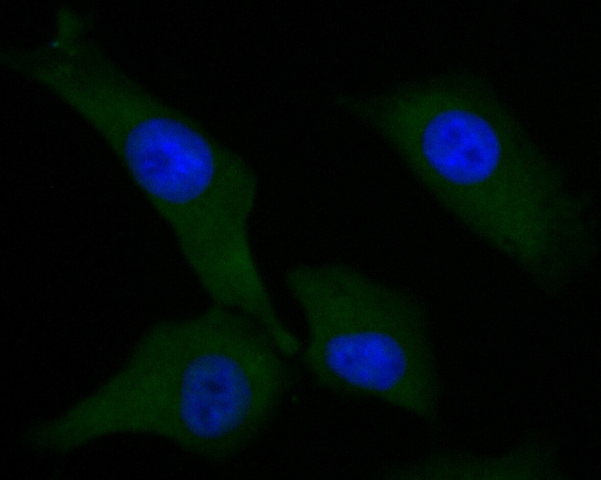
- Immunofluorescent analysis of AKT3 in A549 cells (green). Samples were formalin fixed, permeabilized with 0.1% Triton X-100 in TBS (1 hour, room temperature) and blocked with 1% BSA (15 min, room temperature), incubated with AKT3 polyclonal antibody (Product # PA5-109345) at a dilution of 1:50 (1 hour, room temperature), and followed by Alexa Fluor 488 Goat anti-Rabbit IgG and DAPI (blue) with a dilution of 1:100.
Supportive validation
- Submitted by
- Invitrogen Antibodies (provider)
- Main image
- Experimental details
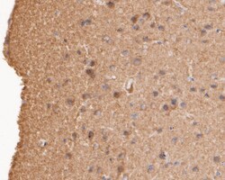
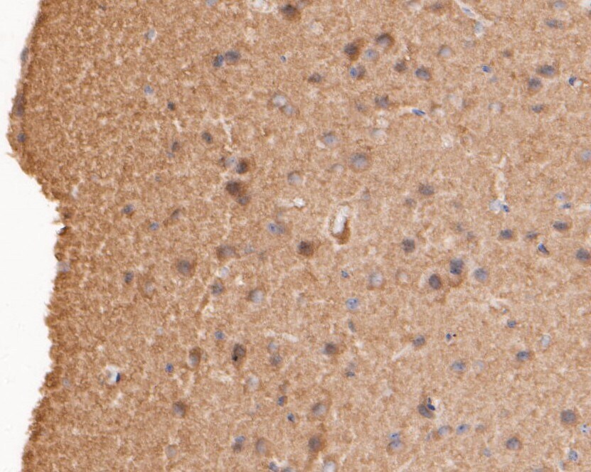
- Immunohistochemistry analysis of AKT3 in paraffin-embedded mouse brain tissue. Samples were heat mediated antigen retrieval with sodium citrate buffer (pH 6.0, 20 min) and blocked in 5% BSA (30 min, room temperature), incubated with AKT3 polyclonal antibody (Product # PA5-109345) at a dilution of 1:200 (30 min, room temperature), and followed by HRP conjugated compact polymer system, DAB, counterstained with hematoxylin (mounted with DPX).
- Submitted by
- Invitrogen Antibodies (provider)
- Main image

- Experimental details
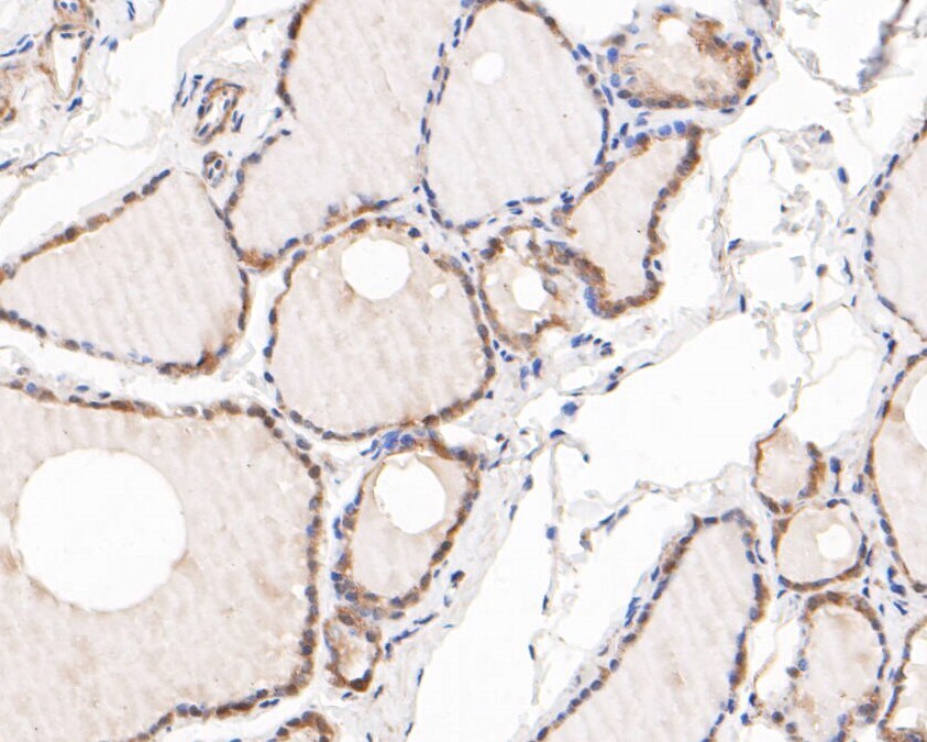
- Immunohistochemistry analysis of AKT3 in paraffin-embedded human thyroid tissue. Samples were heat mediated antigen retrieval with sodium citrate buffer (pH 6.0, 20 min) and blocked in 5% BSA (30 min, room temperature), incubated with AKT3 polyclonal antibody (Product # PA5-109345) at a dilution of 1:200 (30 min, room temperature), and followed by HRP conjugated compact polymer system, DAB, counterstained with hematoxylin (mounted with DPX).
- Submitted by
- Invitrogen Antibodies (provider)
- Main image
- Experimental details
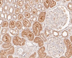
- Immunohistochemistry analysis of AKT3 in paraffin-embedded human kidney tissue. Samples were heat mediated antigen retrieval with sodium citrate buffer (pH 6.0, 20 min) and blocked in 5% BSA (30 min, room temperature), incubated with AKT3 polyclonal antibody (Product # PA5-109345) at a dilution of 1:200 (30 min, room temperature), and followed by HRP conjugated compact polymer system, DAB, counterstained with hematoxylin (mounted with DPX).

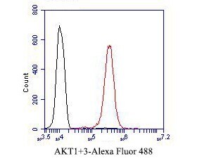
Supportive validation
- Submitted by
- Invitrogen Antibodies (provider)
- Main image
- Experimental details
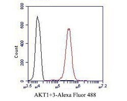
- Flow cytometry of AKT3 in EA.hy926 cells, unlabelled sample (control; cells without incubation with primary antibody; black). Samples were incubated with AKT3 polyclonal antibody (Product # PA5-109345) at a dilution of 1:50, followed by Alexa Fluor 488-conjugated goat anti-rabbit IgG with a dilution of 1:1000 (30 min).