 Explore
 Validate
 Learn
Learn Western blot
 Immunocytochemistry
Antibody data
- Antibody Data
- Antigen structure
- References [1]
- Comments [0]
- Validations
- Immunocytochemistry [2]
- Immunohistochemistry [1]
- Other assay [4]
Submit
Validation data
Reference
Comment
Report error
- Product number
- PA5-29696 - Provider product page

- Provider
- Invitrogen Antibodies
- Product name
- AKT3 Polyclonal Antibody
- Antibody type
- Polyclonal
- Antigen
- Recombinant full-length protein
- Description
- Recommended positive controls: Raji, mouse brain, AKT3-transfected 293T cells. Predicted reactivity: Mouse (100%), Rat (99%), Pig (100%), Rhesus Monkey (100%), Chimpanzee (100%), Bovine (100%). Store product as a concentrated solution. Centrifuge briefly prior to opening the vial.
- Reactivity
- Human, Mouse
- Host
- Rabbit
- Isotype
- IgG
- Vial size
- 100 μL
- Concentration
- 2.15 mg/mL
- Storage
- Store at 4°C short term. For long term storage, store at -20°C, avoiding freeze/thaw cycles.
Submitted references AKT3 deficiency in M2 macrophages impairs cutaneous wound healing by disrupting tissue remodeling.
Gu S, Dai H, Zhao X, Gui C, Gui J
Aging 2020 Apr 14;12(8):6928-6946
Aging 2020 Apr 14;12(8):6928-6946
No comments: Submit comment
Supportive validation
- Submitted by
- Invitrogen Antibodies (provider)
- Main image

- Experimental details
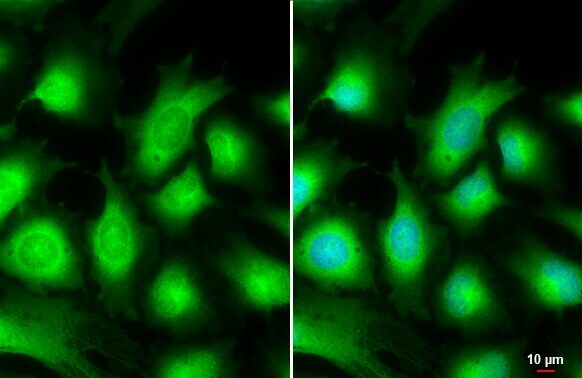
- Immunocytochemistry-Immunofluorescence analysis of AKT3 was performed in HeLa cells fixed in 4% paraformaldehyde at RT for 15 min. Green: AKT3 Polyclonal Antibody (Product # PA5-29696) diluted at 1:500. Blue: Hoechst 33342 staining. Scale bar = 10 µm.
- Submitted by
- Invitrogen Antibodies (provider)
- Main image

- Experimental details
- Immunocytochemistry-Immunofluorescence analysis of AKT3 was performed in HeLa cells fixed in 4% paraformaldehyde at RT for 15 min. Green: AKT3 Polyclonal Antibody (Product # PA5-29696) diluted at 1:500. Blue: Hoechst 33342 staining. Scale bar = 10 µm.

Supportive validation
- Submitted by
- Invitrogen Antibodies (provider)
- Main image

- Experimental details
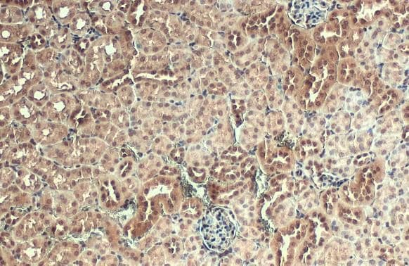
- AKT3 Polyclonal Antibody detects AKT3 protein at nucleus by immunohistochemical analysis. Sample: Paraffin-embedded mouse kidney. AKT3 stained by AKT3 Polyclonal Antibody (Product # PA5-29696) diluted at 1:500. Antigen Retrieval: Citrate buffer, pH 6.0, 15 min.
Supportive validation
- Submitted by
- Invitrogen Antibodies (provider)
- Main image

- Experimental details
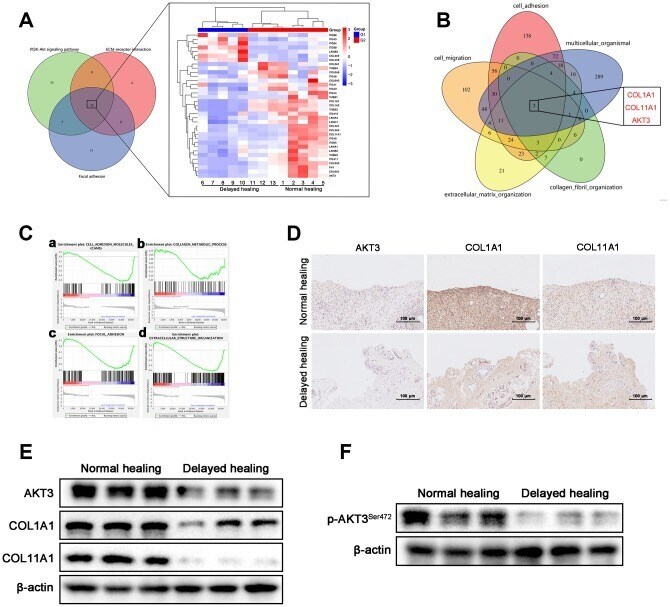
- Figure 3 Downregulation of AKT3, COL1A1, and COL11A1 in delayed cutaneous wound tissue. ( A ) Venn diagram of the KEGG pathway. ( a ) Venn analysis identified 35 genes that were enriched in PI3K-AKT signaling, ECM-receptor interactions, and focal adhesion. ( b ) The heatmap expression profile of the 35 changed genes. ( B ) Venn diagram of GO analysis for the tissue remodeling-associated biological functions. AKT3, COL1A1, and COL11A1 were enriched. ( C a - d ) Gene set enrichment analysis (GSEA) of cutaneous wound tissue. The genes associated with ( a ) cell adhesion molecules, ( b ) collagen metabolic processes, ( c ) focal adhesion, and ( d ) extracellular structural organization were negatively enriched in the delayed cutaneous wound tissue. ( D ) IHC staining of AKT3, COL1A1, and COL11A1 in cutaneous wound tissue (200 x). The levels of all three proteins were reduced in the delayed wound tissue. ( E ) Decreased AKT3, COL1A1, and COL11A1 protein levels in delayed cutaneous wound tissue. ( F ) Total AKT3 and phosphorylated-Ser472 AKT3 levels were decreased in delayed cutaneous wound tissue. All the experiments were repeated at least three times.
- Submitted by
- Invitrogen Antibodies (provider)
- Main image
- Experimental details
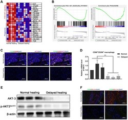
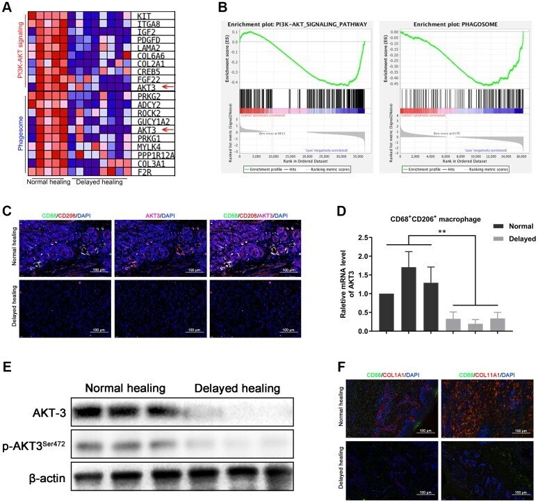
- Figure 4 Loss of AKT3 in M2 macrophages inhibited extracellular COL1A1 and COL11A1 expression. ( A ) GSEA showed that negatively enriched genes were associated with PI3K-AKT signaling and phagosomes in delayed cutaneous wound tissue. ( B ) Heatmap of the top 10 genes related to PI3K-AKT signaling and phagosomes; AKT3 was downregulated in both functional enrichment sets in the delayed cutaneous wound tissue. ( C ) Immunofluorescence of cutaneous wound tissue (n = 6). CD68- (green) and CD206-(red) positive M2 macrophages were reduced in the delayed cutaneous wound tissue. AKT3 (pink) was decreased in the M2 macrophages. ( D ) qRT-PCR showed decreased AKT3 mRNA expression in the delayed cutaneous wound tissue-derived M2 macrophages. ( E ) Western blotting verified the reduction and loss of AKT3 in M2 macrophages from delayed cutaneous wound tissue. ( F ) Immunofluorescence of COL1A1 and COL11A1 in CD68-positive macrophages in cutaneous wound tissue. ( a ) Decreased CD68-positive macrophage infiltration and COL1A1 protein expression were observed in delayed cutaneous wound tissue. ( b ) Decreased COL11A1 protein expression also accompanied the reduced CD68-positive macrophage infiltration. All the experiments were repeated at least three times.
- Submitted by
- Invitrogen Antibodies (provider)
- Main image
- Experimental details
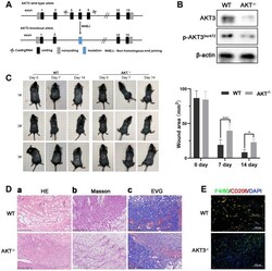
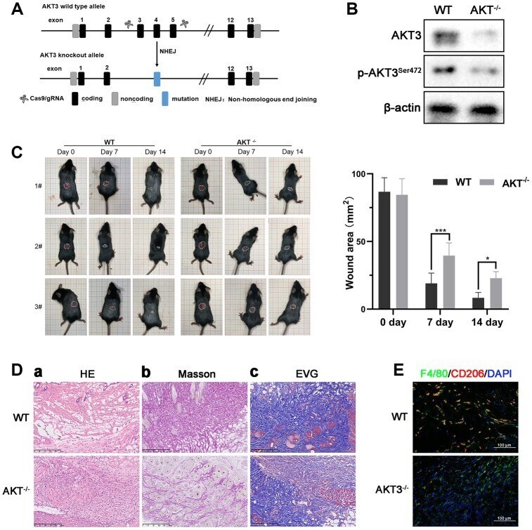
- Figure 6 Loss of AKT3 delayed cutaneous wound healing in mice. ( A ) Schematic of AKT3 knockout in mice. ( B ) Western blotting for AKT3 levels in AKT3 +/+ and AKT3 -/- mice (n = 6). ( C ) AKT3 knockout delayed cutaneous wound healing in mice by days 7 and 14 post-injury. ( D a - c ) Histological staining of mouse cutaneous wound tissue. ( a ) H&E staining showed more inflammatory cells in the wound tissue of AKT3 -/- mice and incomplete tissue integrity (n = 6). ( b ) Masson staining showed the numbers of collagenous and muscular fibers were reduced in the wound tissue of AKT3 -/- mice (n = 6). ( c ) EVG staining showed that number of elastin fibers were decreased in the wound tissue of AKT3 -/- mice (n = 6). ( E ) IF staining showed the F40/80 and CD206 expression in mouse cutaneous wound tissue. All the experiments were repeated at least three times.
- Submitted by
- Invitrogen Antibodies (provider)
- Main image
- Experimental details
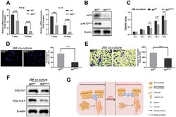
- Figure 7 M2 macrophages from AKT3 -/- mice failed to promote cell proliferation and migration ex vivo. ( A ) TGF-beta and IL-10 mRNA levels were decreased in delayed cutaneous wound tissue 7 th and 14 th day post-injury in mice (n = 3). ( B ) Western blotting demonstrated the loss of AKT3 in M2 macrophages from AKT3 -/- mice. ( C , D ) CCK-8 and EdU assays demonstrated that M2 macrophages from AKT3 -/- mice were incapable of promoting JB6 cell proliferation ( C ) or DNA replication ( D ), respectively. ( E ) Transwell migration assay showed M2 macrophages from AKT3 -/- mice could not promote JB6 cell migration. ( F ) COL1A1 and COL11A1 protein levels in JB6 were not increased by co-culture with M2 macrophages from AKT3 -/- mice. ( G ) The schematic illustration of the role of M2 macrophage AKT3 deficiency in delayed cutaneous wound healing. All the experiments were repeated at least three times.
