 Explore
 Validate
 Learn
Learn Flow cytometry
Antibody data
- Antibody Data
- Antigen structure
- References [1]
- Comments [0]
- Validations
- Flow cytometry [2]
Submit
Validation data
Reference
Comment
Report error
- Product number
- FAB1544P - Provider product page

- Provider
- Novus Biologicals
- Product name
- Goat Polyclonal Insulin R/CD220 Antibody
- Antibody type
- Polyclonal
- Description
- Antigen Affinity-purified. Detects human Insulin R/CD220 in direct ELISAs and Western blots. In direct ELISAs, approximately 20% cross-reactivity with recombinant mouse Insulin R is observed and less than 5% cross-reactivity with recombinant human INSRR is observed.
- Reactivity
- Human, Mouse
- Host
- Goat
- Isotype
- IgG
- Vial size
- 100 Tests
- Storage
- Protect from light. Do not freeze. 12 months from date of receipt, 2 to 8 degreesC as supplied.
Submitted references Co-targeting the IGF system and HIF-1 inhibits migration and invasion by (triple-negative) breast cancer cells.
Mancini M, Gariboldi MB, Taiana E, Bonzi MC, Craparotta I, Pagin M, Monti E
British journal of cancer 2014 Jun 10;110(12):2865-73
British journal of cancer 2014 Jun 10;110(12):2865-73
No comments: Submit comment
Supportive validation
- Submitted by
- Novus Biologicals (provider)
- Main image

- Experimental details
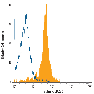
- Detection of Insulin R/CD220 in Human Blood Monocytes by Flow Cytometry. Human peripheral blood monocytes were stained with Goat Anti-Human/Mouse Insulin R/CD220 PE-conjugated Antigen Affinity-purified Polyclonal Antibody (Catalog # FAB1544P, filled histogram) or isotype control antibody (Catalog # IC108P, open histogram). View our protocol for Staining Membrane-associated Proteins.
- Submitted by
- Novus Biologicals (provider)
- Main image
- Experimental details
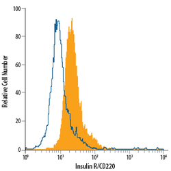
- Detection of Insulin R/CD220 in Neuro-2A Mouse Cell Line by Flow Cytometry. Neuro-2A mouse neuroblastoma cell line was stained with Goat Anti-Human/Mouse Insulin R/CD220 PE-conjugated Antigen Affinity-purified Polyclonal Antibody (Catalog # FAB1544P, filled histogram) or isotype control antibody (Catalog # IC108P, open histogram). View our protocol for Staining Membrane-associated Proteins.
