 Explore
 Validate
 Learn
Learn Western blot
Antibody data
- Antibody Data
- Antigen structure
- References [5]
- Comments [0]
- Validations
- Western blot [1]
- Immunocytochemistry [1]
- Other assay [1]
Submit
Validation data
Reference
Comment
Report error
- Product number
- 44-809G - Provider product page

- Provider
- Invitrogen Antibodies
- Product name
- Phospho-INSR (Tyr1334) Polyclonal Antibody
- Antibody type
- Polyclonal
- Antigen
- Synthetic peptide
- Description
- This antibody has been negatively preadsorbed using a non-phosphopeptide corresponding to the site of phosphorylation to remove antibody that is reactive with non-phosphorylated Insulin Receptor. The final product is generated by affinity chromatography using a Insulin Receptor-derived peptide that is phosphorylated at tyrosine 1334. This antibody recognizes human insulin receptor phosphorylated at tyrosine 1334 as well as the human IR Isoform B phosphorylated at tyrosine 1332. Western blot positive control used was insulin-treated CHO-T cells expressing Human Insulin Receptor.
- Reactivity
- Human, Mouse, Rat
- Host
- Rabbit
- Isotype
- IgG
- Vial size
- 100 µL
- Storage
- -20°C
Submitted references Late-life targeting of the IGF-1 receptor improves healthspan and lifespan in female mice.
Novel Monoclonal Antibody Is an Allosteric Insulin Receptor Antagonist That Induces Insulin Resistance.
Novel method demonstrates differential ligand activation and phosphatase-mediated deactivation of insulin receptor tyrosine-specific phosphorylation.
Specific insulin/IGF1 hybrid receptor activation assay reveals IGF1 as a more potent ligand than insulin.
Leukocyte antigen-related deficiency enhances insulin-like growth factor-1 signaling in vascular smooth muscle cells and promotes neointima formation in response to vascular injury.
Mao K, Quipildor GF, Tabrizian T, Novaj A, Guan F, Walters RO, Delahaye F, Hubbard GB, Ikeno Y, Ejima K, Li P, Allison DB, Salimi-Moosavi H, Beltran PJ, Cohen P, Barzilai N, Huffman DM
Nature communications 2018 Jun 19;9(1):2394
Nature communications 2018 Jun 19;9(1):2394
Novel Monoclonal Antibody Is an Allosteric Insulin Receptor Antagonist That Induces Insulin Resistance.
Cieniewicz AM, Kirchner T, Hinke SA, Nanjunda R, D'Aquino K, Boayke K, Cooper PR, Perkinson R, Chiu ML, Jarantow S, Johnson DL, Whaley JM, Lacy ER, Lingham RB, Liang Y, Kihm AJ
Diabetes 2017 Jan;66(1):206-217
Diabetes 2017 Jan;66(1):206-217
Novel method demonstrates differential ligand activation and phosphatase-mediated deactivation of insulin receptor tyrosine-specific phosphorylation.
Cieniewicz AM, Cooper PR, McGehee J, Lingham RB, Kihm AJ
Cellular signalling 2016 Aug;28(8):1037-47
Cellular signalling 2016 Aug;28(8):1037-47
Specific insulin/IGF1 hybrid receptor activation assay reveals IGF1 as a more potent ligand than insulin.
Slaaby R
Scientific reports 2015 Jan 21;5:7911
Scientific reports 2015 Jan 21;5:7911
Leukocyte antigen-related deficiency enhances insulin-like growth factor-1 signaling in vascular smooth muscle cells and promotes neointima formation in response to vascular injury.
Niu XL, Li J, Hakim ZS, Rojas M, Runge MS, Madamanchi NR
The Journal of biological chemistry 2007 Jul 6;282(27):19808-19
The Journal of biological chemistry 2007 Jul 6;282(27):19808-19
No comments: Submit comment
Supportive validation
- Submitted by
- Invitrogen Antibodies (provider)
- Main image

- Experimental details
- Western blot using IR (pY1334) polyclonal antibody, rabbit

Supportive validation
- Submitted by
- Invitrogen Antibodies (provider)
- Main image
- Experimental details
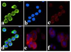
- Immunofluorescence analysis of INSR (pY1334) was done on 70% confluent log phase MCF7 cells with insulin treatment (100nM for 5 min). The cells were fixed with 4% paraformaldehyde for 15 minutes, permeabilized with 0.25% Triton™ X-100 for 10 minutes, and blocked with 5% BSA for 1 hour at room temperature. The cells were labeled with INSR (pY1334) Rabbit polyclonal Antibody (Product # 44-809G) at 1:250 dilution in 1% BSA and incubated for 3 hours at room temperature and then labeled with Alexa Fluor 488 Goat Anti-Rabbit IgG Secondary Antibody (Product # A-11008) at a dilution of 1:400 for 30 minutes at room temperature (Panel a: green). Nuclei (Panel b: blue) were stained with SlowFade® Gold Antifade Mountant DAPI (Product # S36938). F-actin (Panel c: red) was stained with Alexa Fluor 594 Phalloidin (Product # A12381). Panel d is a merged image showing membrane localization. Panel e is untreated MCF7 cells. Panel f shows no primary antibody control. The images were captured at 20X magnification.

Supportive validation
- Submitted by
- Invitrogen Antibodies (provider)
- Main image

- Experimental details
- Figure 1 Specific insulin/IGF1 receptor hybrid activation assay. Western blot analysis of immunoprecipitations (a, b and c) with 24-31 (lanes 1-12) and with 83-7 (lanes 13 and 14) on lysates from BHK cells expressing IR-A and IGF1R (lanes 1-8), IR-A (lanes 9, 10, 13 and 14) or IGF1R (lanes 11 and 12). Cell lysate analysis (d, e and f). The blots were probed with IR pY1334 (a and d), IGF1R N-20 (b and e) and IR C-19 (c and f). Cells were either unstimulated (lanes 1 and 5) or stimulated with 1, 10 or 100 nM insulin (lanes 2, 3 and 4), with IGF1 (lanes 6, 7, and 8), with 100 nM insulin (lanes 9, 11 and 13) or with 100 nM IGF1 (lanes 10, 12 and 14) for 30 minutes at 37degC. The data shown is one representative of four independent experiments. BHK, baby hamster kidney; IGF1, Insulin-like Growth Factor 1; IGF1R, Insulin-like Growth Factor 1 Receptor; IR, Insulin Receptor; IP, ImmunoPrecipitation.
