 Explore
 Validate
 Learn
Learn Western blot
 ELISA
ELISAAntibody data
- Antibody Data
- Antigen structure
- References [0]
- Comments [0]
- Validations
- Western blot [3]
- Immunocytochemistry [2]
Submit
Validation data
Reference
Comment
Report error
- Product number
- AHR0221 - Provider product page

- Provider
- Invitrogen Antibodies
- Product name
- INSR alpha Monoclonal Antibody (83-14)
- Antibody type
- Monoclonal
- Antigen
- Purifed from natural sources
- Description
- This antibody reacts with an epitope at aa 469-592 (exon 7/8). It primarily reacts with human, but also reacts very weakly with cow, pig and sheep. This antibody can inhibit insulin binding (~80%). It can also be used in a Tyrosine Kinase assay (Ab-mediated capture on microtitre plates). Without BSA, this antibody can be used as an insulin-like agonist. Without BSA, it can also be used as both a capture and detection antibody in a sandwich ELISA.
- Reactivity
- Human, Bovine, Porcine
- Host
- Mouse
- Isotype
- IgG
- Antibody clone number
- 83-14
- Vial size
- 500 µL
- Concentration
- 0.2 mg/mL
- Storage
- 4° C
No comments: Submit comment
Supportive validation
- Submitted by
- Invitrogen Antibodies (provider)
- Main image

- Experimental details
- Western blot analysis of Insulin Receptor alpha was performed by loading 25 ug of 293T (lane 1), Hela (lane 2) and PC12 (lane 3) cell lysates onto an SDS polyacrylamide gel. Proteins were transferred to a PVDF membrane and blocked at 4_C overnight. The membrane was probed with a Insulin Receptor alpha monoclonal antibody (Product # AHR0221) at a dilution of 1:200 overnight at 4ÁC, washed in TBST, and probed with an HRP-conjugated secondary antibody for 1 hr at room temperature in the dark. Chemiluminescent detection was performed using Pierce ECL Plus Western Blotting Substrate (Product # 32132). Results show a band at ~95kDa.

- Submitted by
- Invitrogen Antibodies (provider)
- Main image

- Experimental details
- Western blot analysis was performed on membrane enriched extract (30 µg lysate) of IM9 (Lane 1). The blot was probed with Anti-INSR alpha Monoclonal Antibody (Product # AHR0221, 1 µg/mL) and detected by chemiluminescence using Goat anti-Mouse IgG (H+L) Superclonal™ Secondary Antibody, HRP conjugate (Product # A28177, 0.25 µg/mL, 1:4000 dilution). A 130 kDa band corresponding to INSR alpha was observed in the cell line tested.

Supportive validation
- Submitted by
- Invitrogen Antibodies (provider)
- Main image
- Experimental details
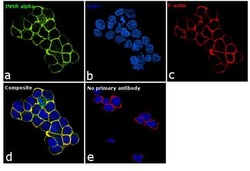
- Immunofluorescence analysis of INSR alpha was performed using log phase IM-9 cells. The cells were fixed with 4% paraformaldehyde for 10 minutes, permeabilized with 0.1% Triton™ X-100 for 10 minutes, and blocked with 1% BSA for 1 hour at room temperature. The cells were labeled with INSR alpha Monoclonal Antibody (83-14) (Product # AHR0221) at 5 µg/mL in 0.1% BSA, incubated overnight at 4 degree and then labeled with Goat anti-Mouse IgG (H+L) Superclonal™ Secondary Antibody, Alexa Fluor® 488 conjugate (Product # A28175) at a dilution of 1:2000 for 45 minutes at room temperature (Panel a: green). Nuclei (Panel b: blue) were stained with SlowFade® Gold Antifade Mountant with DAPI (Product # S36938). F-actin (Panel c: red) was stained with Rhodamine Phalloidin (Product # R415, 1:300). Panel d represents the merged image showing membranous localization. Panel e represents control cells with no primary antibody to assess background. The images were captured at 60X magnification.

- Submitted by
- Invitrogen Antibodies (provider)
- Main image

- Experimental details
- Immunofluorescent analysis of Insulin Receptor alpha (green) showing staining in the membrane of HepG2 cells (right) compared to a negative control without primary antibody (left). Formalin-fixed cells were permeabilized with 0.1% Triton X-100 in TBS for 5-10 minutes and blocked with 3% BSA-PBS for 30 minutes at room temperature. Cells were probed with an Insulin Receptor alpha monoclonal antibody (Product # AHR0221) in 3% BSA-PBS at a dilution of 1:20 and incubated overnight at 4ºC in a humidified chamber. Cells were washed with PBST and incubated with a DyLight-conjugated secondary antibody in PBS at room temperature in the dark. F-actin (red) was stained with a fluorescent red phalloidin and nuclei (blue) were stained with Hoechst or DAPI. Images were taken at a magnification of 60x.
