Explore
 Validate
 Learn
Learn ELISA
ELISA Immunocytochemistry
Antibody data
- Antibody Data
- Antigen structure
- References [3]
- Comments [0]
- Validations
- Immunocytochemistry [2]
- Immunohistochemistry [1]
- Flow cytometry [4]
- Other assay [1]
Submit
Validation data
Reference
Comment
Report error
- Product number
- AHR0231 - Provider product page

- Provider
- Invitrogen Antibodies
- Product name
- INSR alpha Monoclonal Antibody (83-7)
- Antibody type
- Monoclonal
- Antigen
- Purifed from natural sources
- Description
- This antibody is specific for IR and shows no cross-reactivity with insulin-like growth factor (IGF)-receptors. The epitope for this monoclonal antibody is conformational and is located in exon 3. Staining of formalin-fixed, paraffin tissues requires digestion of tissue sections with pepsin at 1mg/mL in Tris-HCl, pH 2.0, for 15 min at room temperature or 10 min at 37ºC. Recommended positive controls include IM-9 lymphocytes, placenta, or breast carcinoma.
- Reactivity
- Human, Bovine, Porcine, Rabbit
- Host
- Mouse
- Isotype
- IgG
- Antibody clone number
- 83-7
- Vial size
- 500 μL
- Concentration
- 0.2 mg/mL
- Storage
- 4°C
Submitted references Regulation of age-associated insulin resistance by MT1-MMP-mediated cleavage of insulin receptor.
Display of Single-Chain Insulin-like Peptides on a Yeast Surface.
Sequential cleavage of insulin receptor by calpain 2 and γ-secretase impairs insulin signalling.
Guo X, Asthana P, Gurung S, Zhang S, Wong SKK, Fallah S, Chow CFW, Che S, Zhai L, Wang Z, Ge X, Jiang Z, Wu J, Zhang Y, Wu X, Xu K, Lin CY, Kwan HY, Lyu A, Zhou Z, Bian ZX, Wong HLX
Nature communications 2022 Jun 29;13(1):3749
Nature communications 2022 Jun 29;13(1):3749
Display of Single-Chain Insulin-like Peptides on a Yeast Surface.
Jeong MY, Rutter J, Chou DH
Biochemistry 2019 Jan 22;58(3):182-188
Biochemistry 2019 Jan 22;58(3):182-188
Sequential cleavage of insulin receptor by calpain 2 and γ-secretase impairs insulin signalling.
Yuasa T, Amo-Shiinoki K, Ishikura S, Takahara M, Matsuoka T, Kaneto H, Kuroda A, Matsuhisa M, Hashida S
Diabetologia 2016 Dec;59(12):2711-2721
Diabetologia 2016 Dec;59(12):2711-2721
No comments: Submit comment
Supportive validation
- Submitted by
- Invitrogen Antibodies (provider)
- Main image
- Experimental details
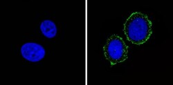
- Immunofluorescent analysis of Insulin Receptor alpha (green) showing staining in the cytoplasm and membrane of MCF-7 cells (right) compared to a negative control without primary antibody (left). Formalin-fixed cells were permeabilized with 0.1% Triton X-100 in TBS for 5-10 minutes and blocked with 3% BSA-PBS for 30 minutes at room temperature. Cells were probed with an Insulin Receptor alpha monoclonal antibody (Product # AHR0231) in 3% BSA-PBS at a dilution of 1:20 and incubated overnight at 4 ºC in a humidified chamber. Cells were washed with PBST and incubated with a DyLight-conjugated secondary antibody in PBS at room temperature in the dark. F-actin (red) was stained with a fluorescent red phalloidin and nuclei (blue) were stained with Hoechst or DAPI. Images were taken at a magnification of 60x.
- Submitted by
- Invitrogen Antibodies (provider)
- Main image

- Experimental details
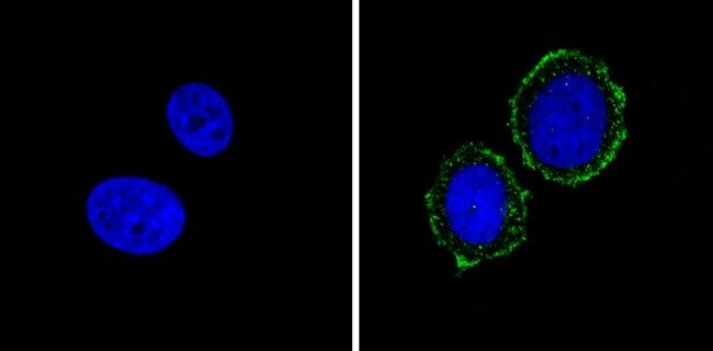
- Immunofluorescent analysis of Insulin Receptor alpha (green) showing staining in the cytoplasm and membrane of MCF-7 cells (right) compared to a negative control without primary antibody (left). Formalin-fixed cells were permeabilized with 0.1% Triton X-100 in TBS for 5-10 minutes and blocked with 3% BSA-PBS for 30 minutes at room temperature. Cells were probed with an Insulin Receptor alpha monoclonal antibody (Product # AHR0231) in 3% BSA-PBS at a dilution of 1:20 and incubated overnight at 4 ºC in a humidified chamber. Cells were washed with PBST and incubated with a DyLight-conjugated secondary antibody in PBS at room temperature in the dark. F-actin (red) was stained with a fluorescent red phalloidin and nuclei (blue) were stained with Hoechst or DAPI. Images were taken at a magnification of 60x.

Supportive validation
- Submitted by
- Invitrogen Antibodies (provider)
- Main image
- Experimental details
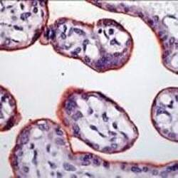
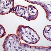
- Immunohistochemical analysis of INSR/Insulin Receptor beta in formalin-fixed, paraffin-embedded human placenta tissue using an INSR monoclonal antibody (Product # AHR0231). Detection was performed with a peroxidase-conjugate and AEC chromogen. Note cell membrane staining of trophoblasts.
Supportive validation
- Submitted by
- Invitrogen Antibodies (provider)
- Main image

- Experimental details
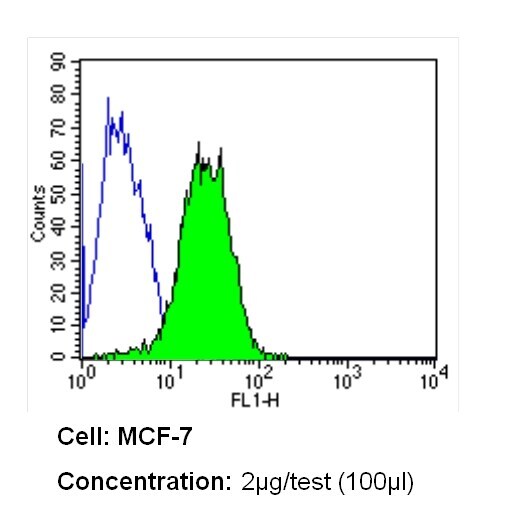
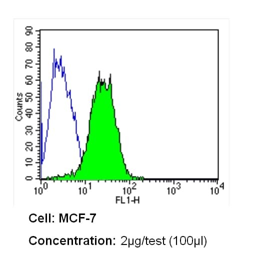
- Flow cytometry analysis of Insulin Receptor alpha in MCF-7 cells (green) compared to an isotype control (blue). Cells were harvested, adjusted to a concentration of 1-5x10^6 cells/mL, fixed with 2% paraformaldehyde and washed with PBS. Cells were blocked with a 2% solution of BSA-PBS for 30 min at room temperature and incubated with a Insulin Receptor alpha monoclonal antibody (Product # AHR0231) at a dilution of 2 µg/test for 60 min at room temperature. Cells were then incubated for 40 min at room temperature in the dark using a Dylight 488-conjugated secondary antibody and re-suspended in PBS for FACS analysis.
- Submitted by
- Invitrogen Antibodies (provider)
- Main image
- Experimental details
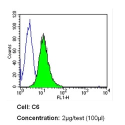
- Flow cytometry analysis of Insulin Receptor alpha in C6 cells (green) compared to an isotype control (blue). Cells were harvested, adjusted to a concentration of 1-5x10^6 cells/mL, fixed with 2% paraformaldehyde and washed with PBS. Cells were blocked with a 2% solution of BSA-PBS for 30 min at room temperature and incubated with a Insulin Receptor alpha monoclonal antibody (Product # AHR0231) at a dilution of 2 µg/test for 60 min at room temperature. Cells were then incubated for 40 min at room temperature in the dark using a Dylight 488-conjugated secondary antibody and re-suspended in PBS for FACS analysis.
- Submitted by
- Invitrogen Antibodies (provider)
- Main image

- Experimental details
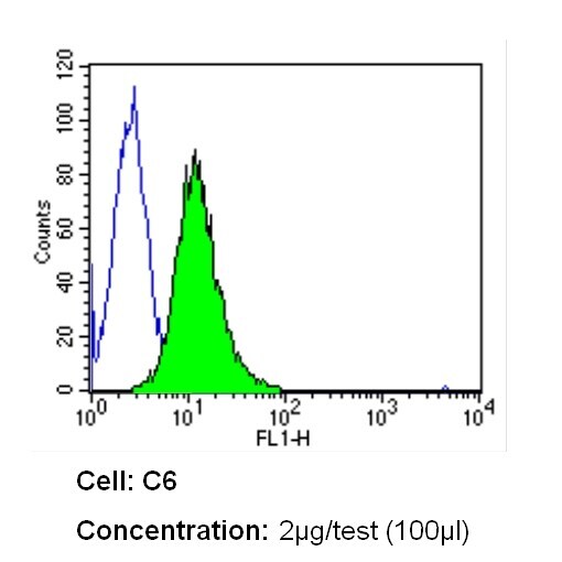
- Flow cytometry analysis of Insulin Receptor alpha in C6 cells (green) compared to an isotype control (blue). Cells were harvested, adjusted to a concentration of 1-5x10^6 cells/mL, fixed with 2% paraformaldehyde and washed with PBS. Cells were blocked with a 2% solution of BSA-PBS for 30 min at room temperature and incubated with a Insulin Receptor alpha monoclonal antibody (Product # AHR0231) at a dilution of 2 µg/test for 60 min at room temperature. Cells were then incubated for 40 min at room temperature in the dark using a Dylight 488-conjugated secondary antibody and re-suspended in PBS for FACS analysis.

- Submitted by
- Invitrogen Antibodies (provider)
- Main image

- Experimental details
- Flow cytometry analysis of Insulin Receptor alpha in MCF-7 cells (green) compared to an isotype control (blue). Cells were harvested, adjusted to a concentration of 1-5x10^6 cells/mL, fixed with 2% paraformaldehyde and washed with PBS. Cells were blocked with a 2% solution of BSA-PBS for 30 min at room temperature and incubated with a Insulin Receptor alpha monoclonal antibody (Product # AHR0231) at a dilution of 2 µg/test for 60 min at room temperature. Cells were then incubated for 40 min at room temperature in the dark using a Dylight 488-conjugated secondary antibody and re-suspended in PBS for FACS analysis.
Supportive validation
- Submitted by
- Invitrogen Antibodies (provider)
- Main image
- Experimental details
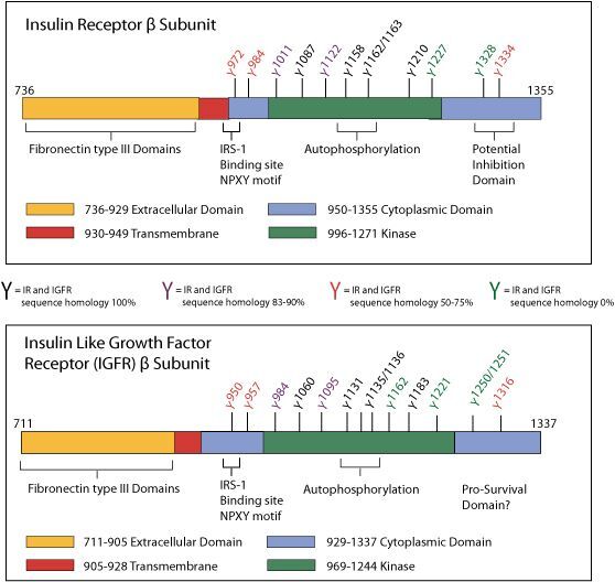
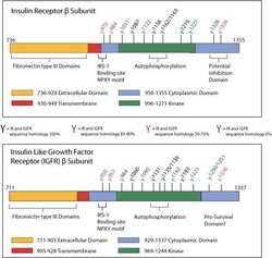
- Insulin Receptor (IR) and Insulin-like Growth Factor-1 Receptor (IGF1R) Protein Schematic-The insulin receptor (IR) is a receptor tyrosine kinase (RTK) that influences multiple signaling pathways through activation of a series of phosphorylation cascades. The IR is a heterotetrameric protein consisting of two ligand-binding a subunits and two b subunits that each contain a tyrosine kinase domain. Upon binding of insulin to the extracellular domain, the receptor is autophosphorylated to activate its intrinsic tyrosine kinase activity. The insulin-like growth factor-1 receptor (IGF1R) is similar to the IR, but exhibits a higher binding affinity for IGF-1 and activates a subset of distinct signaling pathways. Unique phosphorylation sites are said to contribute to signaling differences between IR and IGF1R, and may play a role in the different regulatory functions of the two proteins.