Explore
 Validate
 Learn
Learn Western blot
 ELISA
ELISA Immunocytochemistry
Antibody data
- Antibody Data
- Antigen structure
- References [8]
- Comments [0]
- Validations
- Immunocytochemistry [7]
- Immunohistochemistry [2]
- Other assay [1]
Submit
Validation data
Reference
Comment
Report error
- Product number
- MA5-13783 - Provider product page

- Provider
- Invitrogen Antibodies
- Product name
- INSR Monoclonal Antibody (CT-3)
- Antibody type
- Monoclonal
- Antigen
- Recombinant protein fragment
- Description
- MA5-13783 targets Insulin Receptor beta in ELISA, ICC/IF, IHC (P), Affinity Purification, and WB applications and shows reactivity with Human, mouse, Non-human primate, and Rat samples. The MA5-13783 immunogen is recombinant-fragment including the C-terminal 100 amino acids of human insulin receptor.
- Reactivity
- Human, Mouse
- Host
- Mouse
- Isotype
- IgG
- Antibody clone number
- CT-3
- Vial size
- 500 μL
- Concentration
- 0.2 mg/mL
- Storage
- 4°C
Submitted references Investigating the role of the IGF axis as a predictor of biochemical recurrence in prostate cancer patients post-surgery.
Hyperphosphorylation of Tau induced by naturally secreted amyloid-β at nanomolar concentrations is modulated by insulin-dependent Akt-GSK3β signaling pathway.
Selective response to insulin versus insulin-like growth factor-I and -II and up-regulation of insulin receptor splice variant B in the differentiated mouse mammary epithelium.
Inhibitory effects of nordihydroguaiaretic acid (NDGA) on the IGF-1 receptor and androgen dependent growth of LAPC-4 prostate cancer cells.
Inhibition by transmembrane peptides of chimeric insulin receptors.
Coordinated regulation of insulin signaling by the protein tyrosine phosphatases PTP1B and TCPTP.
The use of protein tyrosine phosphatase 1B and insulin receptor immunostains to differentiate nonalcoholic from alcoholic steatohepatitis in liver biopsy specimens.
Focal overexpression of insulin-like growth factor 2 by hepatocytes and cholangiocytes in viral liver cirrhosis.
Breen KJ, O'Neill A, Murphy L, Fan Y, Boyce S, Fitzgerald N, Dorris E, Brady L, Finn SP, Hayes BD, Treacy A, Barrett C, Aziz MA, Kay EW, Fitzpatrick JM, Watson RWG
The Prostate 2017 Sep;77(12):1288-1300
The Prostate 2017 Sep;77(12):1288-1300
Hyperphosphorylation of Tau induced by naturally secreted amyloid-β at nanomolar concentrations is modulated by insulin-dependent Akt-GSK3β signaling pathway.
Tokutake T, Kasuga K, Yajima R, Sekine Y, Tezuka T, Nishizawa M, Ikeuchi T
The Journal of biological chemistry 2012 Oct 12;287(42):35222-35233
The Journal of biological chemistry 2012 Oct 12;287(42):35222-35233
Selective response to insulin versus insulin-like growth factor-I and -II and up-regulation of insulin receptor splice variant B in the differentiated mouse mammary epithelium.
Berlato C, Doppler W
Endocrinology 2009 Jun;150(6):2924-33
Endocrinology 2009 Jun;150(6):2924-33
Inhibitory effects of nordihydroguaiaretic acid (NDGA) on the IGF-1 receptor and androgen dependent growth of LAPC-4 prostate cancer cells.
Ryan CJ, Zavodovskaya M, Youngren JF, Campbell M, Diamond M, Jones J, Shiry L, Allan G, Maddux BA, Goldfine ID
The Prostate 2008 Aug 1;68(11):1232-40
The Prostate 2008 Aug 1;68(11):1232-40
Inhibition by transmembrane peptides of chimeric insulin receptors.
Bennasroune A, Gardin A, Auzan C, Clauser E, Dirrig-Grosch S, Meira M, Appert-Collin A, Aunis D, Crémel G, Hubert P
Cellular and molecular life sciences : CMLS 2005 Sep;62(18):2124-31
Cellular and molecular life sciences : CMLS 2005 Sep;62(18):2124-31
Coordinated regulation of insulin signaling by the protein tyrosine phosphatases PTP1B and TCPTP.
Galic S, Hauser C, Kahn BB, Haj FG, Neel BG, Tonks NK, Tiganis T
Molecular and cellular biology 2005 Jan;25(2):819-29
Molecular and cellular biology 2005 Jan;25(2):819-29
The use of protein tyrosine phosphatase 1B and insulin receptor immunostains to differentiate nonalcoholic from alcoholic steatohepatitis in liver biopsy specimens.
Sanderson SO, Smyrk TC
American journal of clinical pathology 2005 Apr;123(4):503-9
American journal of clinical pathology 2005 Apr;123(4):503-9
Focal overexpression of insulin-like growth factor 2 by hepatocytes and cholangiocytes in viral liver cirrhosis.
Sedlaczek N, Hasilik A, Neuhaus P, Schuppan D, Herbst H
British journal of cancer 2003 Mar 10;88(5):733-9
British journal of cancer 2003 Mar 10;88(5):733-9
No comments: Submit comment
Supportive validation
- Submitted by
- Invitrogen Antibodies (provider)
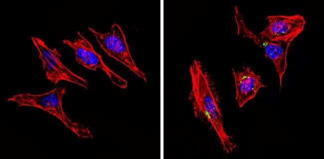
- Main image

- Experimental details
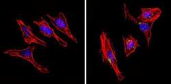
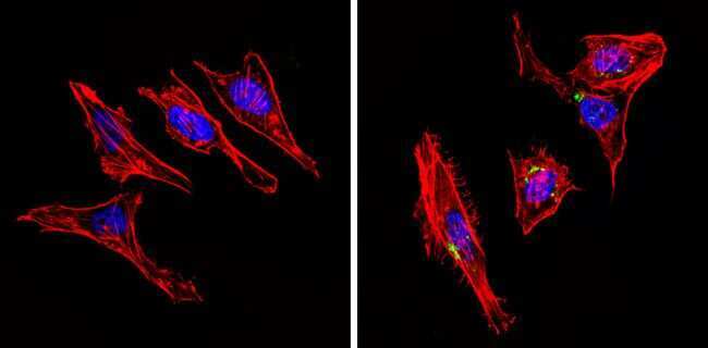
- Immunofluorescent analysis of Insulin Receptor beta (green) showing staining in the cytoplasm of C6 cells (right) compared to a negative control without primary antibody (left). Formalin-fixed cells were permeabilized with 0.1% Triton X-100 in TBS for 5-10 minutes and blocked with 3% BSA-PBS for 30 minutes at room temperature. Cells were probed with an Insulin Receptor beta monoclonal antibody (Product # MA5-13783) in 3% BSA-PBS at a dilution of 1:20 and incubated overnight at 4 ºC in a humidified chamber. Cells were washed with PBST and incubated with a DyLight-conjugated secondary antibody in PBS at room temperature in the dark. F-actin (red) was stained with a fluorescent red phalloidin and nuclei (blue) were stained with Hoechst or DAPI. Images were taken at a magnification of 60x.
- Submitted by
- Invitrogen Antibodies (provider)
- Main image
- Experimental details
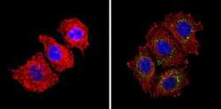
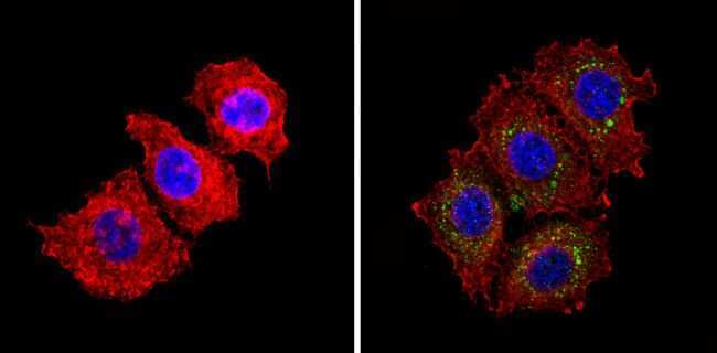
- Immunofluorescent analysis of Insulin Receptor beta (green) showing staining in the cytoplasm of Hela cells (right) compared to a negative control without primary antibody (left). Formalin-fixed cells were permeabilized with 0.1% Triton X-100 in TBS for 5-10 minutes and blocked with 3% BSA-PBS for 30 minutes at room temperature. Cells were probed with an Insulin Receptor beta monoclonal antibody (Product # MA5-13783) in 3% BSA-PBS at a dilution of 1:20 and incubated overnight at 4 ºC in a humidified chamber. Cells were washed with PBST and incubated with a DyLight-conjugated secondary antibody in PBS at room temperature in the dark. F-actin (red) was stained with a fluorescent red phalloidin and nuclei (blue) were stained with Hoechst or DAPI. Images were taken at a magnification of 60x.
- Submitted by
- Invitrogen Antibodies (provider)
- Main image
- Experimental details
- Immunofluorescent analysis of Insulin Receptor beta (green) showing staining in the cytoplasm of MCF-7 cells (right) compared to a negative control without primary antibody (left). Formalin-fixed cells were permeabilized with 0.1% Triton X-100 in TBS for 5-10 minutes and blocked with 3% BSA-PBS for 30 minutes at room temperature. Cells were probed with an Insulin Receptor beta monoclonal antibody (Product # MA5-13783) in 3% BSA-PBS at a dilution of 1:20 and incubated overnight at 4 ºC in a humidified chamber. Cells were washed with PBST and incubated with a DyLight-conjugated secondary antibody in PBS at room temperature in the dark. F-actin (red) was stained with a fluorescent red phalloidin and nuclei (blue) were stained with Hoechst or DAPI. Images were taken at a magnification of 60x.
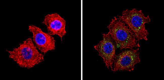
- Submitted by
- Invitrogen Antibodies (provider)
- Main image
- Experimental details
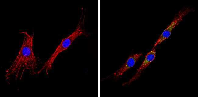
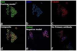
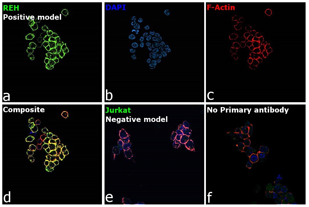
- Immunofluorescence analysis of Insulin receptor was performed using 70% confluent log phase Reh cells. The cells were fixed with 4% paraformaldehyde for 10 minutes, permeabilized with 0.1% Triton™ X-100 for 15 minutes, and blocked with 2% BSA for 45 minutes at room temperature. The cells were labeled with INSR Monoclonal Antibody (CT-3) (Product # MA5-13783) at 1:100 dilution in 0.1% BSA, incubated at 4 degree celsius overnight and then labeled with Donkey anti-Mouse IgG (H+L) Highly Cross-Adsorbed Secondary Antibody, Alexa Fluor Plus 488 (Product # A32766), (1:2000), for 45 minutes at room temperature (Panel a: Green). Nuclei (Panel b: Blue) were stained with ProLong™ Diamond Antifade Mountant with DAPI (Product # P36962). F-actin (Panel c: Red) was stained withRhodamine Phalloidin (Product # R415, 1:300). Panel d represents the merged image showing membrane localization. Panel e represents Jurkat. Panel f represents control cells with no primary antibody to assess background. The images were captured at 60x magnification.
- Submitted by
- Invitrogen Antibodies (provider)
- Main image

- Experimental details
- Immunofluorescent analysis of Insulin Receptor beta (green) showing staining in the cytoplasm of Hela cells (right) compared to a negative control without primary antibody (left). Formalin-fixed cells were permeabilized with 0.1% Triton X-100 in TBS for 5-10 minutes and blocked with 3% BSA-PBS for 30 minutes at room temperature. Cells were probed with an Insulin Receptor beta monoclonal antibody (Product # MA5-13783) in 3% BSA-PBS at a dilution of 1:20 and incubated overnight at 4 ºC in a humidified chamber. Cells were washed with PBST and incubated with a DyLight-conjugated secondary antibody in PBS at room temperature in the dark. F-actin (red) was stained with a fluorescent red phalloidin and nuclei (blue) were stained with Hoechst or DAPI. Images were taken at a magnification of 60x.
- Submitted by
- Invitrogen Antibodies (provider)
- Main image

- Experimental details
- Immunofluorescent analysis of Insulin Receptor beta (green) showing staining in the cytoplasm of MCF-7 cells (right) compared to a negative control without primary antibody (left). Formalin-fixed cells were permeabilized with 0.1% Triton X-100 in TBS for 5-10 minutes and blocked with 3% BSA-PBS for 30 minutes at room temperature. Cells were probed with an Insulin Receptor beta monoclonal antibody (Product # MA5-13783) in 3% BSA-PBS at a dilution of 1:20 and incubated overnight at 4 ºC in a humidified chamber. Cells were washed with PBST and incubated with a DyLight-conjugated secondary antibody in PBS at room temperature in the dark. F-actin (red) was stained with a fluorescent red phalloidin and nuclei (blue) were stained with Hoechst or DAPI. Images were taken at a magnification of 60x.
- Submitted by
- Invitrogen Antibodies (provider)
- Main image

- Experimental details
- Immunofluorescence analysis of Insulin receptor was performed using 70% confluent log phase Reh cells. The cells were fixed with 4% paraformaldehyde for 10 minutes, permeabilized with 0.1% Triton™ X-100 for 15 minutes, and blocked with 2% BSA for 45 minutes at room temperature. The cells were labeled with INSR Monoclonal Antibody (CT-3) (Product # MA5-13783) at 1:100 dilution in 0.1% BSA, incubated at 4 degree celsius overnight and then labeled with Donkey anti-Mouse IgG (H+L) Highly Cross-Adsorbed Secondary Antibody, Alexa Fluor Plus 488 (Product # A32766), (1:2000), for 45 minutes at room temperature (Panel a: Green). Nuclei (Panel b: Blue) were stained with ProLong™ Diamond Antifade Mountant with DAPI (Product # P36962). F-actin (Panel c: Red) was stained withRhodamine Phalloidin (Product # R415, 1:300). Panel d represents the merged image showing membrane localization. Panel e represents Jurkat. Panel f represents control cells with no primary antibody to assess background. The images were captured at 60x magnification.

Supportive validation
- Submitted by
- Invitrogen Antibodies (provider)
- Main image
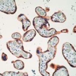
- Experimental details
- Formalin-fixed, paraffin-embedded human placenta stained with Insulin Receptor antibody using peroxidase-conjugate and AEC chromogen. Note cell membrane staining of trophoblasts.
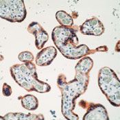
- Submitted by
- Invitrogen Antibodies (provider)
- Main image
- Experimental details
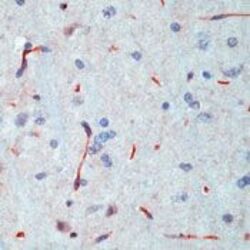
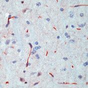
- Formalin-fixed, paraffin-embedded rat brain stained with Insulin Receptor antibody using peroxidase-conjugate and AEC chromogen. Note staining of endothelial cells of capillaries.
Supportive validation
- Submitted by
- Invitrogen Antibodies (provider)
- Main image

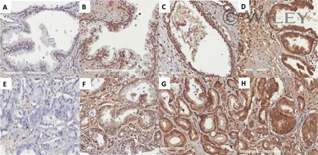
- Experimental details
- Representative images (x20 magnification) of IHC staining of TMA for INSR, 1:50 dilution, (A-D) benign tissue scoring intensity 0-3, (E-H) cancerous tissue scoring intensity 0-3. TMA sections were incubated with primary antibody at optimized concentrations overnight at 4degC. All images taken using Aperio Slide Scanner