Explore
 Validate
 Learn
Learn Western blot
 ELISA
ELISA Immunocytochemistry
Antibody data
- Antibody Data
- Antigen structure
- References [1]
- Comments [0]
- Validations
- Immunocytochemistry [2]
- Immunohistochemistry [2]
- Flow cytometry [2]
- Other assay [1]
Submit
Validation data
Reference
Comment
Report error
- Product number
- MA5-17052 - Provider product page

- Provider
- Invitrogen Antibodies
- Product name
- CDK2 Monoclonal Antibody (1A6)
- Antibody type
- Monoclonal
- Antigen
- Purifed from natural sources
- Description
- MA5-17052 targets CDK2 in indirect ELISA, FACS, ICC, IHC, IF and WB applications and shows reactivity with Human and Mouse samples. The MA5-17052 immunogen is purified recombinant fragment of human CDK2 expressed in E. Coli. MA5-17052 detects CDK2 which has a predicted molecular weight of approximately 33.9kDa.
- Reactivity
- Human, Mouse
- Host
- Mouse
- Isotype
- IgG
- Antibody clone number
- 1A6
- Vial size
- 100 μg
- Concentration
- 1 mg/mL
- Storage
- Store at 4°C short term. For long term storage, store at -20°C, avoiding freeze/thaw cycles.
Submitted references MM-129 as a Novel Inhibitor Targeting PI3K/AKT/mTOR and PD-L1 in Colorectal Cancer.
Hermanowicz JM, Pawlak K, Sieklucka B, Czarnomysy R, Kwiatkowska I, Kazberuk A, Surazynski A, Mojzych M, Pawlak D
Cancers 2021 Jun 26;13(13)
Cancers 2021 Jun 26;13(13)
No comments: Submit comment
Supportive validation
- Submitted by
- Invitrogen Antibodies (provider)
- Main image
- Experimental details
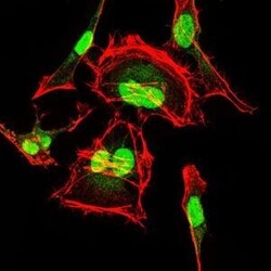
- Immunofluorescence analysis of HeLa cells using CDK2 monoclonal antibody (Product # MA5-17052) (Green). Red: actin filaments have been labeled with phalloidin.

- Submitted by
- Invitrogen Antibodies (provider)
- Main image
- Experimental details
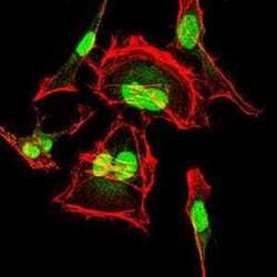
- Immunofluorescence analysis of HeLa cells using CDK2 monoclonal antibody (Product # MA5-17052) (Green). Red: actin filaments have been labeled with phalloidin.

Supportive validation
- Submitted by
- Invitrogen Antibodies (provider)
- Main image

- Experimental details
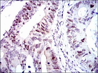
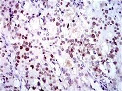
- Immunohistochemical analysis of paraffin-embedded colon cancer tissues using CDK2 monoclonal antibody (Product # MA5-17052) followed with DAB staining.
- Submitted by
- Invitrogen Antibodies (provider)
- Main image
- Experimental details
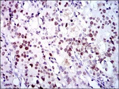
- Immunohistochemical analysis of paraffin-embedded cervical cancer tissues using CDK2 monoclonal antibody (Product # MA5-17052) followed with DAB staining.
Supportive validation
- Submitted by
- Invitrogen Antibodies (provider)
- Main image
- Experimental details
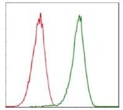
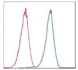
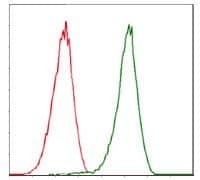
- Flow cytometric analysis of Jurkat cells using CDK2 monoclonal antibody (Product # MA5-17052) (green) and negative control (red).
- Submitted by
- Invitrogen Antibodies (provider)
- Main image
- Experimental details
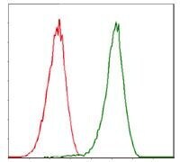
- Flow cytometric analysis of Jurkat cells using CDK2 monoclonal antibody (Product # MA5-17052) (green) and negative control (red).
Supportive validation
- Submitted by
- Invitrogen Antibodies (provider)
- Main image
- Experimental details
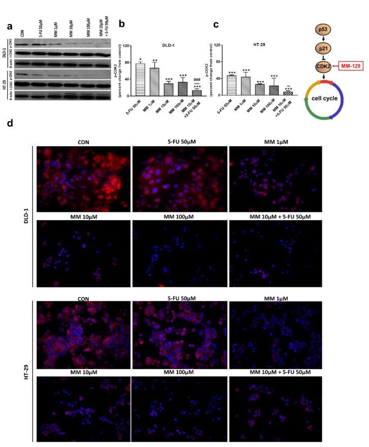
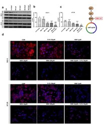
- Figure 8 Phosphorylated CDK2 (p-CDK2), total CDK2 (t-CDK2), and beta-actin expression as determined by Western blot ( a ) and phosphorylated CDK2 (p-CDK2) determined by confocal microscopy ( d ) in DLD-1 and HT-29 cells treated with 5-FU (5-FU 50 uM), MM-129 (MM 1 uM, 10 uM, 100 uM), and their combination (MM 10 uM + 5-FU 50 uM) for 24 h. The samples used for electrophoresis consisted of 20 ug of protein from 6 pooled cell extracts from independent experiments ( n = 6). Band staining was quantified by densitometry ( b , c ). The corresponding uncropped blots are shown in Supplementary Figures S9a-c and S10a-c . Cells were incubated with rabbit polyclonal antibody against phospho CDK2 and secondary goat polyclonal antibody against rabbit (red label). The nuclei were stained with Hoechst 33342 (blue label) ( d ). The results are presented as means +- SDs. * p < 0.05, ** p < 0.01, *** p < 0.001 vs. CON, ^^ p < 0.01 vs. 5-FU, ^^^ p < 0.001 vs. 5-FU, ### p < 0.001 vs. MM-129 at dose 10 uM.