 Explore
 Validate
 Learn
Learn Western blot
Antibody data
- Antibody Data
- Antigen structure
- References [0]
- Comments [0]
- Validations
- Western blot [3]
- Immunocytochemistry [1]
- Immunohistochemistry [1]
Submit
Validation data
Reference
Comment
Report error
- Product number
- PA5-32340 - Provider product page

- Provider
- Invitrogen Antibodies
- Product name
- CDK2 Polyclonal Antibody
- Antibody type
- Polyclonal
- Antigen
- Synthetic peptide
- Description
- Heat-mediated antigen retrieval is recommended prior to staining, using an EDTA buffer for 10 minutes followed by cooling at room temperature for 20 min. Following antigen retrieval, incubate samples with primary antibody for 30 min at room temperature. A suggested positive control is HeLa cell lysate.
- Reactivity
- Human, Mouse
- Host
- Rabbit
- Isotype
- IgG
- Vial size
- 500 µL
- Storage
- Store at 4°C short term. For long term storage, store at -20°C, avoiding freeze/thaw cycles.
No comments: Submit comment
Supportive validation
- Submitted by
- Invitrogen Antibodies (provider)
- Main image
- Experimental details
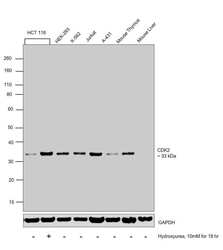
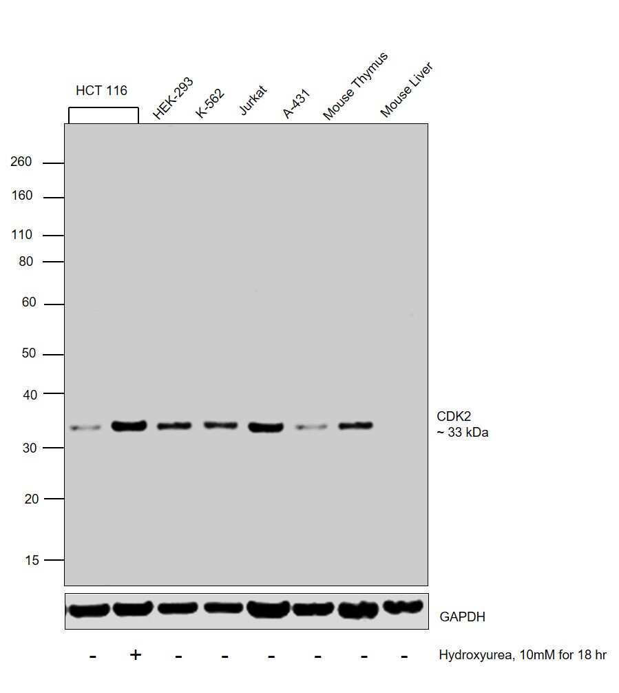
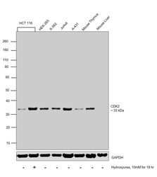
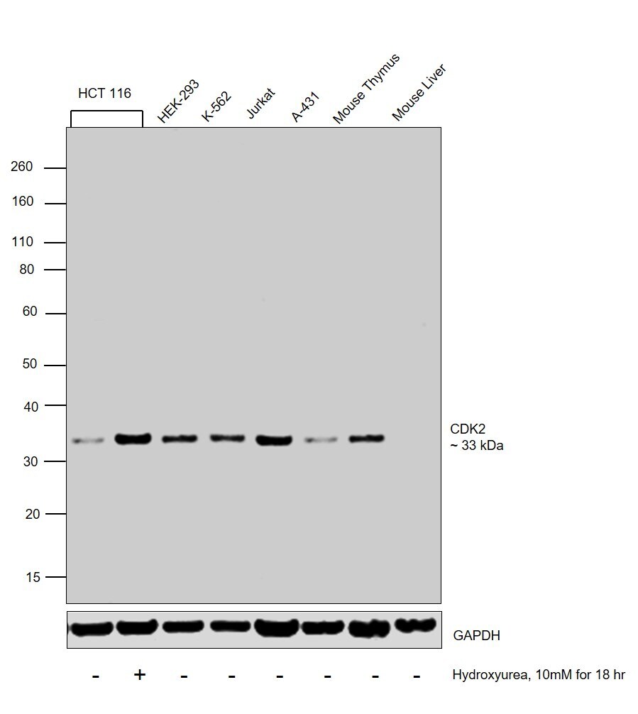
- Western blot was performed using Anti-CDK2 Polyclonal Antibody (Product # PA5-32340) and 33 kDa band corresponding to CDK2 was observed across the cell lines and tissues tested except Mouse Liver which is reported to be negative. Also, the band was induced in HCT 116 upon Hydroxyurea treatment. Whole cell extracts (30 µg lysate) of HCT 116 (Lane 1), HCT 116 treated with Hydroxyurea (10mM for 18 hours) (Lane 2), HEK-293 (Lane 3), K-562 (Lane 4), Jurkat (Lane 5) and A-431 (Lane 6), tissue extracts (30 µg lysate) of Mouse Thymus (Lane 7) and Mouse Liver (Lane 8) were electrophoresed using Novex® NuPAGE® 4-12 % Bis-Tris gel (Product # NP0322BOX). Resolved proteins were then transferred onto a nitrocellulose membrane (Product # IB23001) by iBlot® 2 Dry Blotting System (Product # IB21001). The blot was probed with the primary antibody (1:25 dilution) and detected by chemiluminescence with Goat anti-Rabbit IgG (H+L), Superclonal™ Recombinant Secondary Antibody, HRP (Product # A27036, 1:4000 dilution) using the iBright FL 1000 (Product # A32752). Chemiluminescent detection was performed using Novex® ECL Chemiluminescent Substrate Reagent Kit (Product # WP20005).
- Submitted by
- Invitrogen Antibodies (provider)
- Main image
- Experimental details
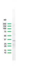
- Western blot analysis of Hela Cells using anti-CDK2 Polyclonal Antibody (Product # PA5-32340). The recommened dilution for this antibody in western blot applications is 1:25.

- Submitted by
- Invitrogen Antibodies (provider)
- Main image
- Experimental details
- Western blot was performed using Anti-CDK2 Polyclonal Antibody (Product # PA5-32340) and 33 kDa band corresponding to CDK2 was observed across the cell lines and tissues tested except Mouse Liver which is reported to be negative. Also, the band was induced in HCT 116 upon Hydroxyurea treatment. Whole cell extracts (30 µg lysate) of HCT 116 (Lane 1), HCT 116 treated with Hydroxyurea (10mM for 18 hours) (Lane 2), HEK-293 (Lane 3), K-562 (Lane 4), Jurkat (Lane 5) and A-431 (Lane 6), tissue extracts (30 µg lysate) of Mouse Thymus (Lane 7) and Mouse Liver (Lane 8) were electrophoresed using Novex® NuPAGE® 4-12 % Bis-Tris gel (Product # NP0322BOX). Resolved proteins were then transferred onto a nitrocellulose membrane (Product # IB23001) by iBlot® 2 Dry Blotting System (Product # IB21001). The blot was probed with the primary antibody (1:25 dilution) and detected by chemiluminescence with Goat anti-Rabbit IgG (H+L), Superclonal™ Recombinant Secondary Antibody, HRP (Product # A27036, 1:4000 dilution) using the iBright FL 1000 (Product # A32752). Chemiluminescent detection was performed using Novex® ECL Chemiluminescent Substrate Reagent Kit (Product # WP20005).
Supportive validation
- Submitted by
- Invitrogen Antibodies (provider)
- Main image
- Experimental details
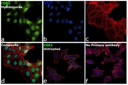
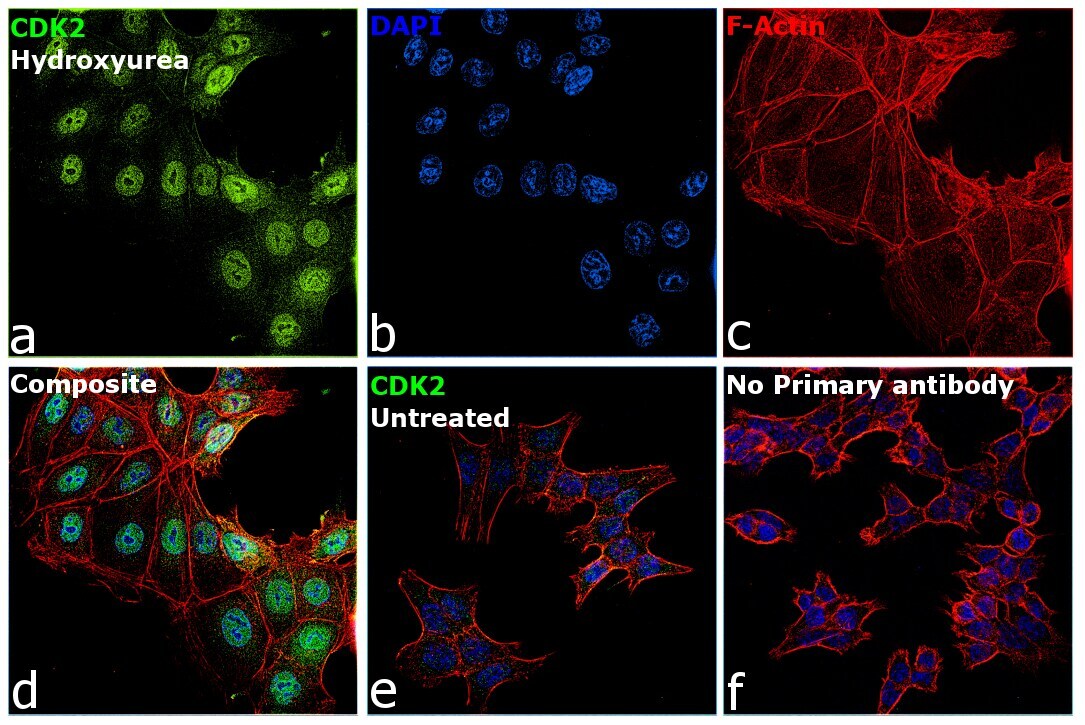
- Immunofluorescence analysis of CDK2 was performed using 70% confluent log phase HCT 116 cells treated with Hydroxyurea (10mM for 18 hours). The cells were fixed with 4% paraformaldehyde for 10 minutes, permeabilized with 0.1% Triton™ X-100 for 15 minutes, and blocked with 2% BSA for 1 hour at room temperature. The cells were labeled with CDK2 Polyclonal Antibody (Product # PA5-32340) at 1:100 dilution in 0.1% BSA, incubated at 4 degree Celsius overnight and then labeled with Goat anti-Rabbit IgG (H+L), Superclonal™ Recombinant Secondary Antibody, Alexa Fluor 488 (Product # A27034, 1:2000 dilution) for 45 minutes at room temperature (Panel a: Green). Nuclei (Panel b: Blue) were stained with SlowFade® Gold Antifade Mountant with DAPI (Product # S36938). F-actin (Panel c: red) was stained with Rhodamine Phalloidin (Product # R415, 1:300). Panel d represents the merged image showing increased CDK2 expression and localization to Nucleus upon treatment with Hydroxyurea. Panel e shows untreated cells with lower expression of CD2. Panel f represents control cells with no primary antibody to assess background. The images were captured at 60X magnification.
Supportive validation
- Submitted by
- Invitrogen Antibodies (provider)
- Main image
- Experimental details
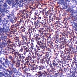
- Immunohistochemical analysis of CDK2 using anti-CDK2 Polyclonal Antibody (Product # PA5-32340) in Tonsil Tissue. The recommened dilution for this antibody in immunohistochemistry applications is 1:100.
