Explore
 Validate
 Learn
Learn Western blot
Antibody data
- Antibody Data
- Antigen structure
- References [3]
- Comments [0]
- Validations
- Western blot [5]
- Immunocytochemistry [5]
- Immunohistochemistry [4]
- Flow cytometry [2]
Submit
Validation data
Reference
Comment
Report error
- Product number
- NBP2-29940 - Provider product page

- Provider
- Novus Biologicals
- Product name
- Rabbit Polyclonal Glutaminase Antibody
- Antibody type
- Polyclonal
- Description
- Protein A purified.
- Reactivity
- Human, Mouse, Rat
- Host
- Rabbit
- Isotype
- IgG
- Vial size
- 0.4 ml
- Concentration
- 0.34 mg/ml
- Storage
- Store at 4C short term. Aliquot and store at -20C long term. Avoid freeze-thaw cycles.
Submitted references NRH:quinone oxidoreductase 2 (NQO2) and glutaminase (GLS) both play a role in large extracellular vesicles (LEV) formation in preclinical LNCaP-C4-2B prostate cancer model of progressive metastasis.
Energy Metabolism Drugs Block Triple Negative Breast Metastatic Cancer Cell Phenotype.
Mitochondrial pyruvate carrier function determines cell stemness and metabolic reprogramming in cancer cells.
Dorai T, Shah A, Summers F, Mathew R, Huang J, Hsieh TC, Wu JM
The Prostate 2018 Nov;78(15):1181-1195
The Prostate 2018 Nov;78(15):1181-1195
Energy Metabolism Drugs Block Triple Negative Breast Metastatic Cancer Cell Phenotype.
Pacheco-Velázquez SC, Robledo-Cadena DX, Hernández-Reséndiz I, Gallardo-Pérez JC, Moreno-Sánchez R, Rodríguez-Enríquez S
Molecular pharmaceutics 2018 Jun 4;15(6):2151-2164
Molecular pharmaceutics 2018 Jun 4;15(6):2151-2164
Mitochondrial pyruvate carrier function determines cell stemness and metabolic reprogramming in cancer cells.
Li X, Han G, Li X, Kan Q, Fan Z, Li Y, Ji Y, Zhao J, Zhang M, Grigalavicius M, Berge V, Goscinski MA, Nesland JM, Suo Z
Oncotarget 2017 Jul 11;8(28):46363-46380
Oncotarget 2017 Jul 11;8(28):46363-46380
No comments: Submit comment
Supportive validation
- Submitted by
- Novus Biologicals (provider)
- Main image
- Experimental details
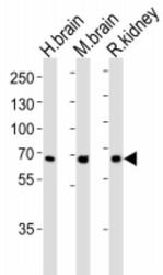
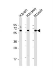
- Western Blot: Glutaminase Antibody [NBP2-29940] - Analysis of lysates from human brain, mouse brain ad rat kidney tissue lysate (from left to right), using (C-term)(Cat. # NBP2-29940). The antibody was diluted at 1:1000 at each lane. A goat anti-rabbit IgG H&L(HRP) at 1:5000 dilution was used as the secondary antibody. Lysates at 35 ug per lane.

- Submitted by
- Novus Biologicals (provider)
- Main image
- Experimental details
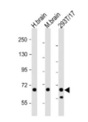
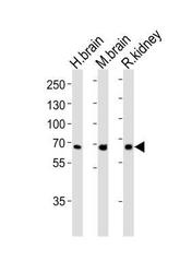
- Western Blot: Glutaminase Antibody [NBP2-29940] - All lanes: Anti-GLS Antibody (C-term) at 1:2000. Lane 1: human brain lysate. Lane 2: mouse brain lysate. Lane 3: 293T/17 whole cell lysate. 20 ug per lane. Secondary Goat Anti-Rabbit IgG, (H+L), Peroxidase conjugated at 1:10000. Predicted band size: 73 kDa. Blocking/Dilution buffer: 5% NFDM/TBST.
- Submitted by
- Novus Biologicals (provider)
- Main image
- Experimental details
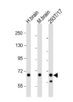
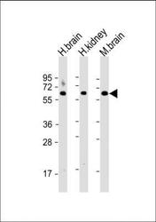
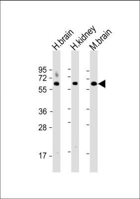
- Western Blot: Glutaminase Antibody [NBP2-29940] - All lanes: Anti-GLS Antibody (C-term) at 1:2000. Lane 1: human brain lysate. Lane 2: human kidney lysate. Lane 3: mouse brain lysate. 20 ug per lane. Secondary Goat Anti-Rabbit IgG, (H+L), Peroxidase conjugated at 1:10000. Predicted band size: 73 kDa. Blocking/Dilution buffer: 5% NFDM/TBST.

- Submitted by
- Novus Biologicals (provider)
- Main image
- Experimental details
- Western Blot: Glutaminase Antibody (RB22875) [NBP2-29940] - Western blot analysis of lysates from human brain, mouse brain and rat kidney tissue lysate (from left to right), using GLS Antibody (C-term)(NBP2-29940). A goat anti-rabbit IgG H&L(HRP) was used as the secondary antibody. Lysates at 35ug per lane.

- Submitted by
- Novus Biologicals (provider)
- Main image
- Experimental details
- Western Blot: Glutaminase Antibody (RB22875) [NBP2-29940] - All lanes : Anti-GLS Antibody (C-term). Lane 1: human brain lysate; Lane 2: human kidney lysate; Lane 3: mouse brain lysate. Lysates/proteins at 20 ug per lane. Secondary Goat Anti-Rabbit IgG, (H+L), Peroxidase conjugated. Predicted band size : 73 kDa. Blocking/Dilution buffer: 5% NFDM/TBST.
Supportive validation
- Submitted by
- Novus Biologicals (provider)
- Main image
- Experimental details
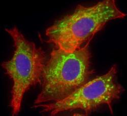
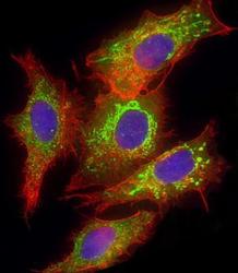
- Immunocytochemistry/Immunofluorescence: Glutaminase Antibody [NBP2-29940] - Analysis of 4% paraformaldehyde-fixed, 0.1% Triton X-100 permeabilized HepG2 cells labeling GLS with antibody at 1:25, followed by DyLight 488-conjugated goat anti-rabbit IgG secondary antibody at 1:200 dilution (green). Immunofluorescence image showing mitochondrion staining on HepG2 cell line. Cytoplasmic actin is detected with DyLight 554 Phalloidin at 1:100 (red). The nuclear counter stain is DAPI (blue).
- Submitted by
- Novus Biologicals (provider)
- Main image

- Experimental details
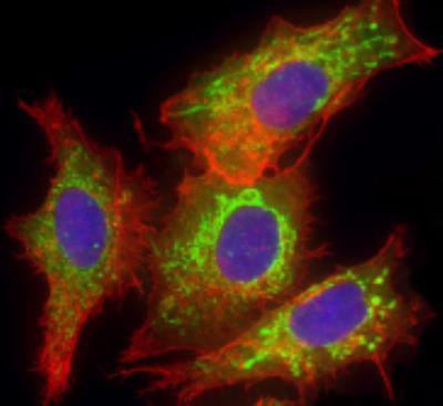
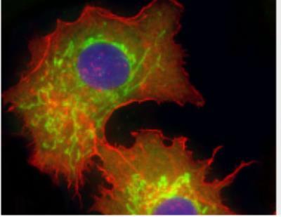
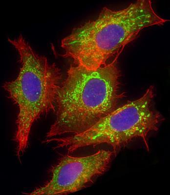
- Immunocytochemistry/Immunofluorescence: Glutaminase Antibody (RB22875) [NBP2-29940] - Immunofluorescent analysis of 4% paraformaldehyde-fixed, 0. 1% Triton X-100 permeabilized U-251 MG cells labeling GLS with NBP2-29940, followed by Dylight(R) 488-conjugated goat anti-Rabbit IgG secondary antibody (green). Immunofluorescence image showing Cytoplasm staining on U-251 MG cell line. Cytoplasmic actin is detected with Dylight(R) 554 Phalloidin(red). The nuclear counter stain is DAPI (blue).
- Submitted by
- Novus Biologicals (provider)
- Main image
- Experimental details
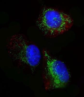
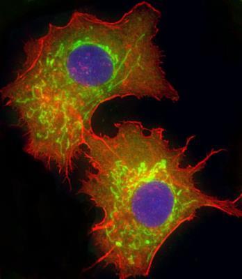
- Immunocytochemistry/Immunofluorescence: Glutaminase Antibody (RB22875) [NBP2-29940] - Immunofluorescent analysis of 4% paraformaldehyde-fixed, 0.1% Triton X-100 permeabilized HepG2 (human liver hepatocellular carcinoma cell line) cells labeling GLS with NBP2-29940, followed by Dylight(R) 488-conjugated goat anti-rabbit IgG secondary antibody (green). Immunofluorescence image showing mitochondrion staining on HepG2 cell line. Cytoplasmic actin is detected with Dylight(R) 554 Phalloidin (red). The nuclear counter stain is DAPI (blue).
- Submitted by
- Novus Biologicals (provider)
- Main image

- Experimental details
- Immunocytochemistry/Immunofluorescence: Glutaminase Antibody (RB22875) [NBP2-29940] - Immunofluorescent analysis of 4% paraformaldehyde-fixed, 0.1% Triton X-100 permeabilized HepG2 (human liver hepatocellular carcinoma cell line) cells labeling GLS with NBP2-29940, followed by Dylight(R) 488-conjugated goat anti-rabbit IgG secondary antibody (green). Immunofluorescence image showing mitochondrion staining on HepG2 cell line. Cytoplasmic actin is detected with Dylight(R) 554 Phalloidin (red). The nuclear counter stain is DAPI (blue).
Supportive validation
- Submitted by
- Novus Biologicals (provider)
- Main image

- Experimental details
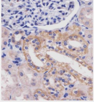
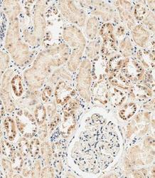
- Immunohistochemistry-Paraffin: Glutaminase Antibody [NBP2-29940] - Staining GLS in human kidney FFPE tissue sections. Tissue was fixed with formaldehyde and blocked with 3% BSA for 0. 5 hour at room temperature. Antigen retrieval by heat mediation with a citrate buffer (pH 6). Samples were incubated with primary antibody (1:25) for 1 hours at 37C. An undiluted biotinylated goat polyvalent antibody was used as the secondary antibody.
- Submitted by
- Novus Biologicals (provider)
- Main image
- Experimental details
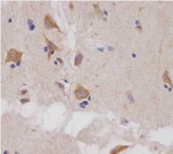
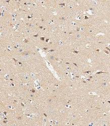
- Immunohistochemistry-Paraffin: Glutaminase Antibody [NBP2-29940] - Staining GLS in human brain FFPE tissue sections. Antigen retrieval by heat mediation with a citrate buffer (pH 6). Samples were incubated with primary antibody (1:25) for 1 hours at 37C. An undiluted biotinylated goat polyvalent antibody was used as the secondary antibody.
- Submitted by
- Novus Biologicals (provider)
- Main image
- Experimental details
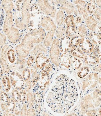
- Immunohistochemistry-Paraffin: Glutaminase Antibody (RB22875) [NBP2-29940] - Immunohistochemical analysis of paraffin-embedded Human kidney tissue using NBP2-22940 performed on the Leica(R) BOND RXm. Tissue was fixed with formaldehyde at room temperature, antigen retrieval was by heat mediation with a EDTA buffer (pH9. 0). Samples were incubated with primary antibody for 1 hr at RT. A undiluted biotinylated CRF Anti-Polyvalent HRP Polymer antibody was used as the secondary antibody.
- Submitted by
- Novus Biologicals (provider)
- Main image
- Experimental details
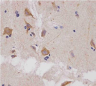
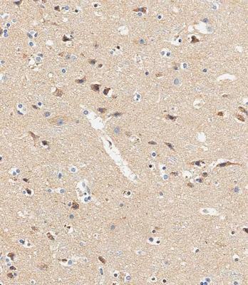
- Immunohistochemistry-Paraffin: Glutaminase Antibody (RB22875) [NBP2-29940] - Immunohistochemical analysis of paraffin-embedded Human brain tissue using NBP2-29940 performed on the Leica(R) BOND RXm. Tissue was fixed with formaldehyde at RT, antigen retrieval was by heat mediation with a EDTA buffer (pH9. 0). Samples were incubated with primary antibody for 1 hr at RT. A undiluted biotinylated CRF Anti-Polyvalent HRP Polymer antibody was used as the secondary antibody.
Supportive validation
- Submitted by
- Novus Biologicals (provider)
- Main image
- Experimental details
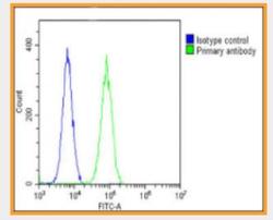
- Flow Cytometry: Glutaminase Antibody [NBP2-29940] - The cells were fixed with 2% paraformaldehyde (10 min) and then permeabilized with 90% methanol for 10 min. The cells were then icubated in 2% bovine serum albumin to block non-specific protein-protein interactions followed by the antibody (1:25 dilution) for 60 min at 37C. The secondary antibody used was Goat-Anti-Rabbit IgG, DyLight 488 Conjugated Highly Cross-Adsorbed at 1:200 dilution for 40 min at 37C. Isotype control antibody (blue line) was rabbit IgG1 (1ug/1x10^6 cells) used under the same conditions. Acquisition of >10, 000 events was performed
- Submitted by
- Novus Biologicals (provider)
- Main image

- Experimental details
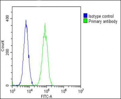
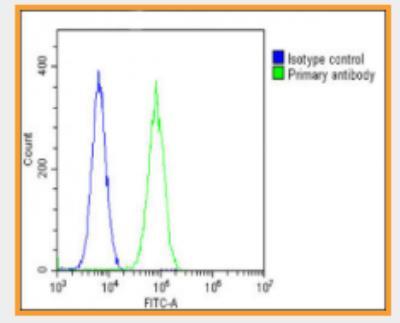
- Flow Cytometry: Glutaminase Antibody (RB22875) [NBP2-29940] - Overlay histogram showing U-2 OS cells stained with NBP2-29940 (green line). The cells were fixed with 2% paraformaldehyde (10 min) and then permeabilized with 90% methanol for 10 min. The cells were then icubated in 2% bovine serum albumin to block non-specific protein-protein interactions followed by the antibody for 60 min at 37C. The secondary antibody used was Goat-Anti-Rabbit IgG, DyLight(R) 488 Conjugated Highly Cross-Adsorbed for 40 min at 37C. Isotype control antibody (blue line) was rabbit IgG1 (1ug/1x10^6 cells) used under the same conditions. Acquisition of >10, 000 events was performed.