 Explore
 Validate
 Learn
Learn Western blot
 Immunocytochemistry
Antibody data
- Antibody Data
- Antigen structure
- References [1]
- Comments [0]
- Validations
- Western blot [3]
- Immunocytochemistry [1]
- Immunoprecipitation [1]
- Immunohistochemistry [3]
Submit
Validation data
Reference
Comment
Report error
- Product number
- GTX131263 - Provider product page

- Provider
- GeneTex
- Product name
- Glutaminase C antibody
- Antibody type
- Polyclonal
- Reactivity
- Human, Mouse
- Host
- Rabbit
Submitted references Metformin Impairs Glutamine Metabolism and Autophagy in Tumour Cells.
Saladini S, Aventaggiato M, Barreca F, Morgante E, Sansone L, Russo MA, Tafani M
Cells 2019 Jan 14;8(1)
Cells 2019 Jan 14;8(1)
No comments: Submit comment
Enhanced validation
Supportive validation
- Submitted by
- GeneTex (provider)
- Enhanced method
- Genetic validation
- Main image
- Experimental details
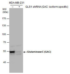
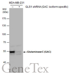
- Glutaminase C (GAC) antibody validation by shRNA knock-down. Non-transfected (-) and GLS1(GAC isoform specific) shRNA-transfected MDA-MB-231 whole cell extracts (30 £gg) were separated by 7.5% SDS-PAGE, and the membrane was blotted with Glutaminase C (GAC) antibody (GTX131263) at a dilution of 1:5000.
Supportive validation
- Submitted by
- GeneTex (provider)
- Main image
- Experimental details
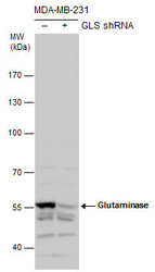
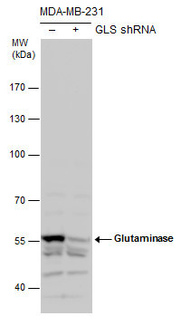
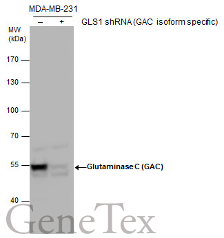
- Glutaminase antibody validation by shRNA knock-down. Non-transfected (-) and Glutaminase shRNA-transfected MDA-MB-231 whole cell extracts (30 ?g) were separated by 7.5% SDS-PAGE, and the membrane was blotted with Glutaminase antibody (GTX131263) diluted by 1:5000.
- Validation comment
- WB
- Submitted by
- GeneTex (provider)
- Main image
- Experimental details
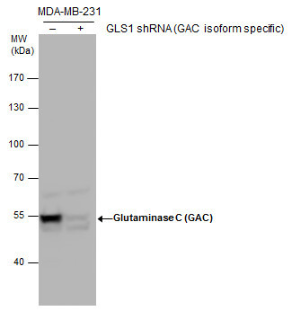
- Glutaminase C (GAC) antibody validation by shRNA knock-down. Non-transfected (-) and GLS1(GAC isoform specific) shRNA-transfected MDA-MB-231 whole cell extracts (30 £gg) were separated by 7.5% SDS-PAGE, and the membrane was blotted with Glutaminase C (GAC) antibody (GTX131263) at a dilution of 1:5000.
Supportive validation
- Submitted by
- GeneTex (provider)
- Main image
- Experimental details
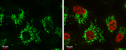
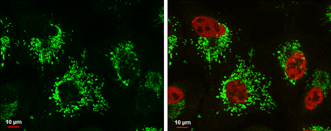
- Glutaminase C antibody detects Glutaminase C protein at mitochondria by immunofluorescent analysis.Sample: A549 cells were fixed in 4% paraformaldehyde at RT for 15 min.Green: Glutaminase C protein stained by Glutaminase C antibody (GTX131263) diluted at 1:500.Red: Histone H3K9ac (acetyl Lys9), a nucleus marker, stained by Histone H3K9ac (acetyl Lys9) antibody [GT464] (GTX630554) diluted at 1:500.Blue: Hoechst 33342 staining.Scale bar = 10 £gm.
Supportive validation
- Submitted by
- GeneTex (provider)
- Main image
- Experimental details
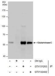
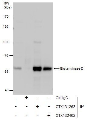
- Immunoprecipitation of Glutaminase C protein from MDA-MB-231 whole cell extracts using 5 £gg of Glutaminase C antibody (GTX131263) or Glutaminase C antibody (GTX132402).Western blot analysis was performed using Glutaminase C antibody (GTX131263).EasyBlot anti-Rabbit IgG (GTX221666-01) was used as a secondary reagent.
Supportive validation
- Submitted by
- GeneTex (provider)
- Main image
- Experimental details
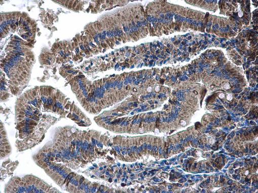
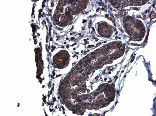
- Glutaminase C antibody detects Glutaminase C protein at cytoplasm on mouse duidenum by immunohistochemical analysis. Sample: Paraffin-embedded mouse duidenum. Glutaminase C antibody (GTX131263) diluted at 1:500.
- Submitted by
- GeneTex (provider)
- Main image
- Experimental details
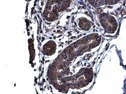
- Glutaminase C antibody detects Glutaminase C protein at cytoplasm on human breast carcinoma by immunohistochemical analysis. Sample: Paraffin-embedded human breast carcinoma. Glutaminase C antibody (GTX131263) diluted at 1:500.
- Submitted by
- GeneTex (provider)
- Main image
- Experimental details
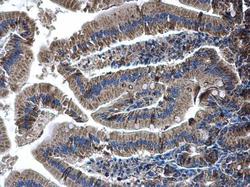
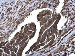
- Glutaminase C antibody detects Glutaminase C protein at cytoplasm on human colon carcinoma by immunohistochemical analysis. Sample: Paraffin-embedded human colon carcinoma. Glutaminase C antibody (GTX131263) diluted at 1:500.
