Explore
 Validate
 Learn
Learn Western blot
Antibody data
- Antibody Data
- Antigen structure
- References [0]
- Comments [0]
- Validations
- Western blot [6]
- Immunocytochemistry [3]
- Immunohistochemistry [2]
- Flow cytometry [1]
Submit
Validation data
Reference
Comment
Report error
- Product number
- ARG55696 - Provider product page

- Provider
- Arigo
- Product name
- anti-Glutaminase antibody
- Antibody type
- Polyclonal
- Antigen
- KLH-conjugated synthetic peptide around aa. 516-545 (C-terminus) of Human Glutaminase.
- Description
- Purification with Protein A and immunogen peptide.
- Reactivity
- Human, Mouse, Rat
- Host
- Rabbit
- Isotype
- IgG
- Vial size
- 100 µl
- Storage
- For continuous use, store undiluted antibody at 2-8°C for up to a week. For long-term storage, aliquot and store at -20°C or below. Storage in frost free freezers is not recommended. Avoid repeated freeze/thaw cycles. Suggest spin the vial prior to opening.
- Handling
- The antibody solution should be gently mixed before use.
No comments: Submit comment
Supportive validation
- Submitted by
- Arigo (provider)
- Main image
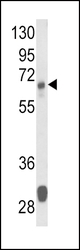
- Experimental details
- Western blot: 35 µg of Mouse liver tissue lysates stained with ARG55696 anti-Glutaminase antibody.
- Submitted by
- Arigo (provider)
- Main image
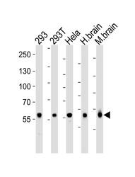
- Experimental details
- Western blot: 35 µg of 293, 293T, HeLa cell line , huamn brain and Mouse brain tissue lysate (from left to right) stained with ARG55696 anti-Glutaminase antibody at 1:1000 dilution.

- Submitted by
- Arigo (provider)
- Main image
- Experimental details
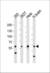
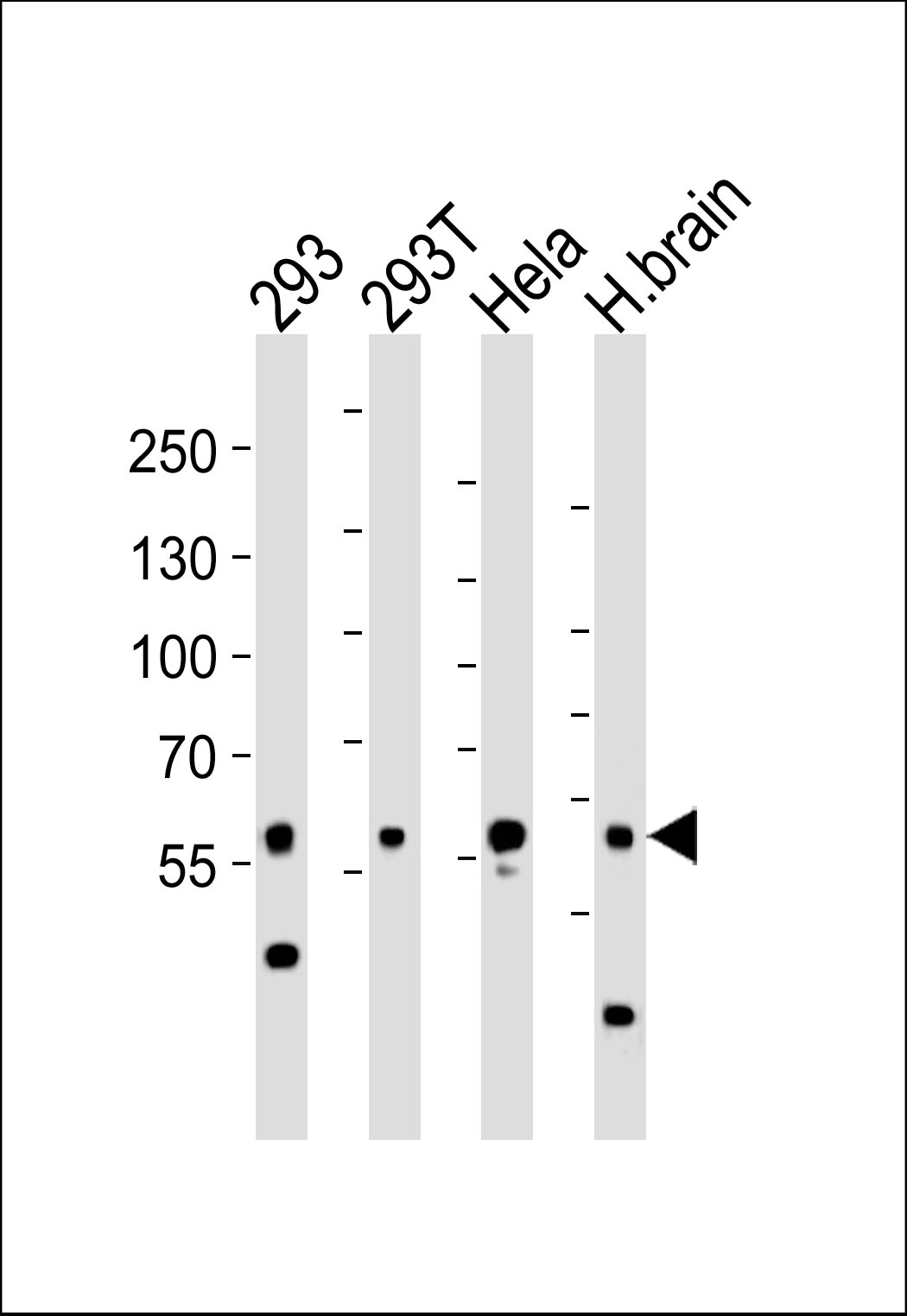
- Western blot: 35 µg of 293, 293T, HeLa cell line and Human brain tissue lysate (from left to right) stained with ARG55696 anti-Glutaminase antibody at 1:1000 dilution.
- Submitted by
- Arigo (provider)
- Main image

- Experimental details
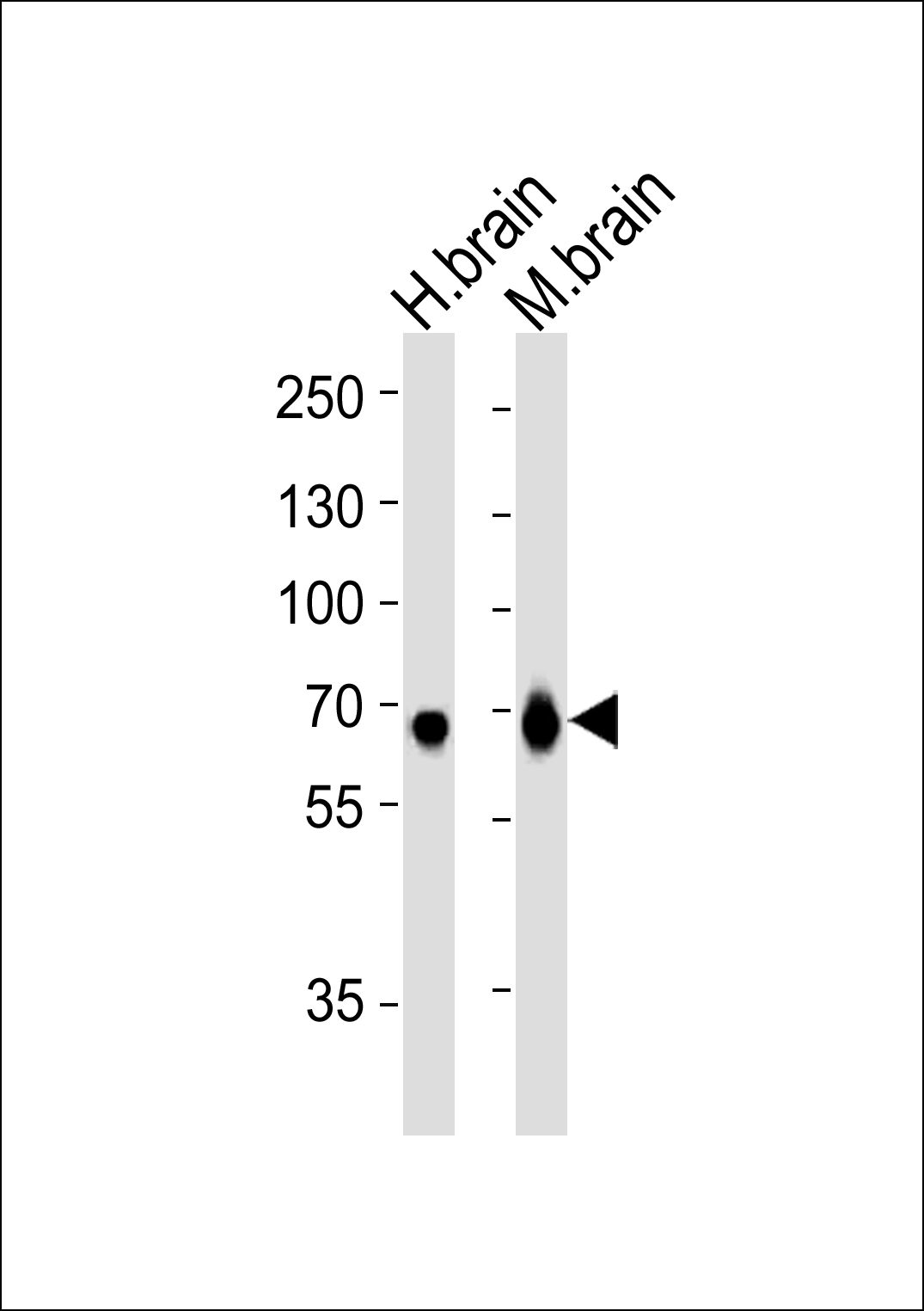
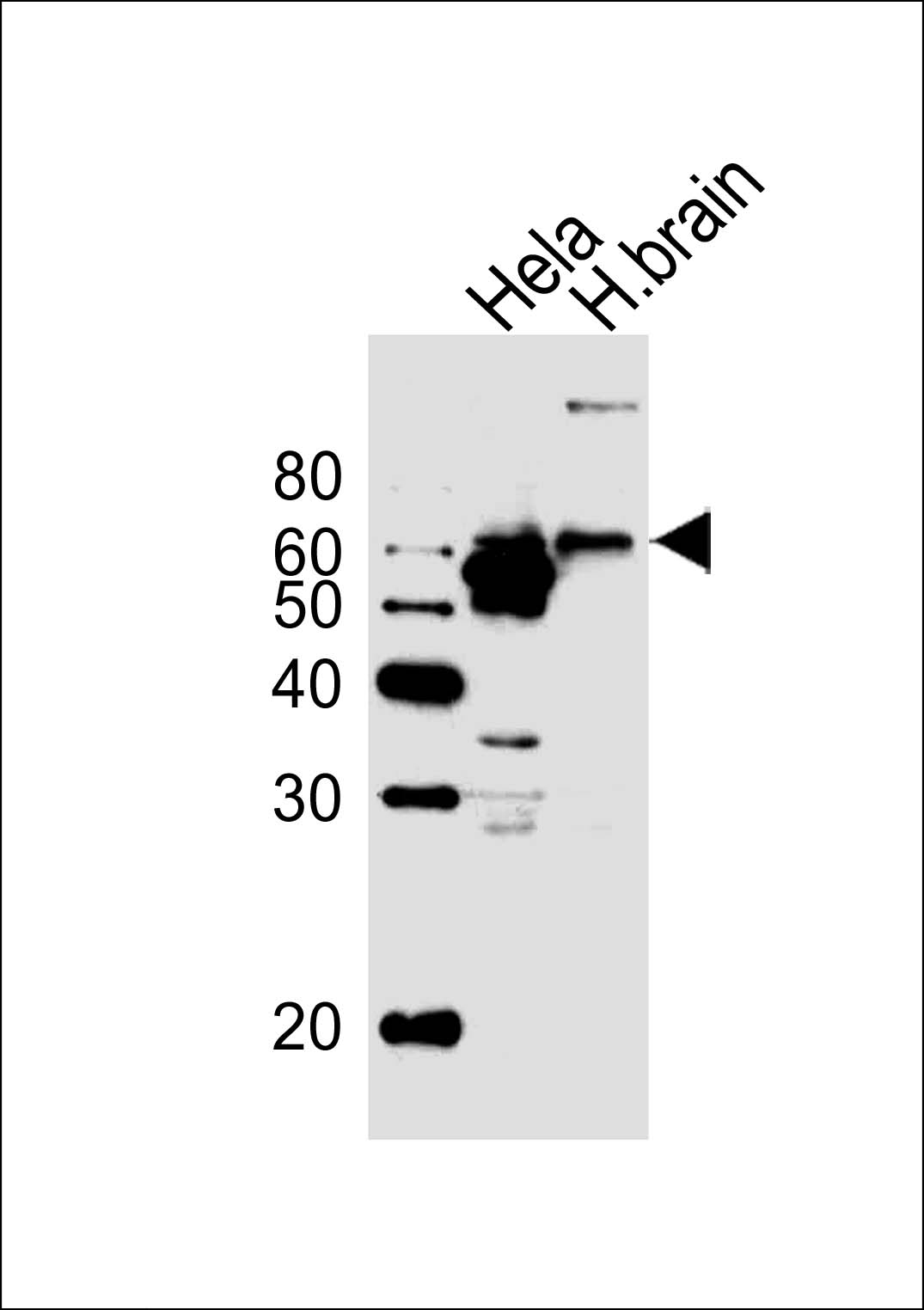
- Western blot: 35 µg of Human brain and Mouse brain tissue lysate (from left to right) stained with ARG55696 anti-Glutaminase antibody at 1:1000 dilution.
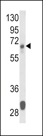
- Submitted by
- Arigo (provider)
- Main image
- Experimental details
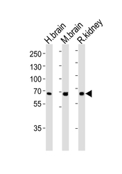
- Western blot: 35 µg of Human brain, Mouse brain ad Rat kidney tissue lysate (from left to right) stained with ARG55696 anti-Glutaminase antibody at 1:1000 dilution.

- Submitted by
- Arigo (provider)
- Main image

- Experimental details
- Western blot: 20 µg of Human brain tissue and HeLa cell line (from left to right) stained with ARG55696 anti-Glutaminase antibody at 1:1000 dilution.
Supportive validation
- Submitted by
- Arigo (provider)
- Main image

- Experimental details
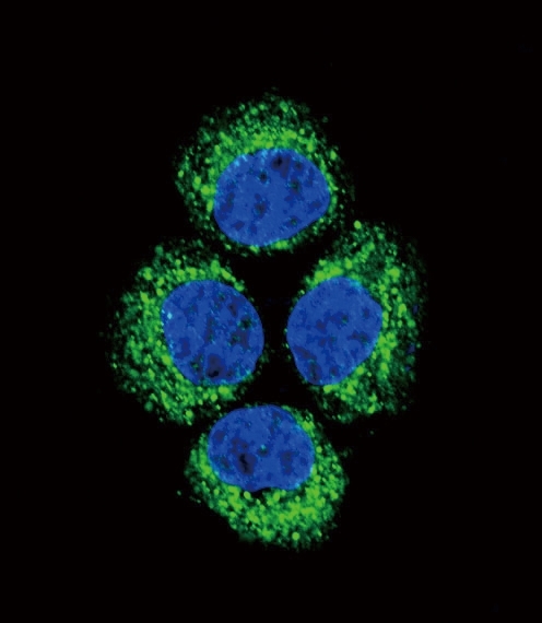
- Immunofluorescence: HeLa cell stained with ARG55696 anti-Glutaminase antibody (green). DAPI was used to stain the cell nuclear (blue).
- Submitted by
- Arigo (provider)
- Main image
- Experimental details
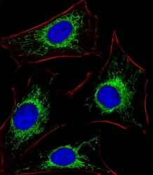
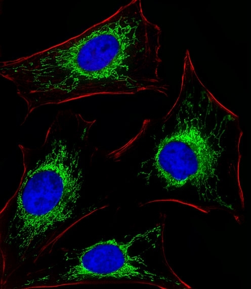
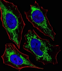
- Immunofluorescence: HeLa cells stained with ARG55696 anti-Glutaminase antibody (green) at 1:25 dilution. DAPI was used to stain the cell nuclear (blue). Cytoplasmic actin was counterstained with Alexa Fluor? 555 conjugated with Phalloidin (red).
- Submitted by
- Arigo (provider)
- Main image
- Experimental details
- Immunofluorescence: HeLa cells stained with ARG55696 anti-Glutaminase antibody (green) at 1:25 dilution. DAPI was used to stain the cell nuclear (blue). Cytoplasmic actin was counterstained with Alexa Fluor? 555 conjugated with Phalloidin (red).

Supportive validation
- Submitted by
- Arigo (provider)
- Main image
- Experimental details
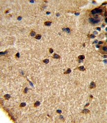
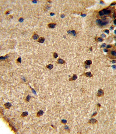
- Immunohistochemistry: formalin fixed and paraffin embedded Mouse brain tissue stained with ARG55696 anti-Glutaminase antibody.
- Submitted by
- Arigo (provider)
- Main image

- Experimental details
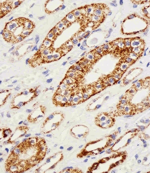
- Immunohistochemistry: paraffin-embedded H.kidney section stained with ARG55696 anti-Glutaminase antibody at 1:25 dilution.
Supportive validation
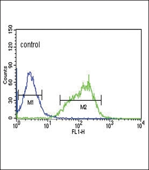
- Submitted by
- Arigo (provider)
- Main image
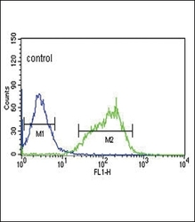
- Experimental details
- Flow Cytometry: HepG2 cells stained with ARG55696 anti-Glutaminase antibody (right histogram) or without primary antibody control (left histogram), followed by incubation with FITC labelled secondary antibody.