 Explore
 Validate
 Learn
Learn Western blot
 Immunocytochemistry
Antibody data
- Antibody Data
- Antigen structure
- References [2]
- Comments [0]
- Validations
- Immunocytochemistry [4]
- Flow cytometry [1]
- Other assay [1]
Submit
Validation data
Reference
Comment
Report error
- Product number
- 701965 - Provider product page

- Provider
- Invitrogen Antibodies
- Product name
- Glutaminase Recombinant Rabbit Monoclonal Antibody (6H5L15)
- Antibody type
- Monoclonal
- Antigen
- Other
- Description
- This antibody is predicted to react with Monkey, Rabbit, Rat and Mouse. Recombinant rabbit monoclonal antibodies are produced using in vitro expression systems. The expression systems are developed by cloning in the specific antibody DNA sequences from immunoreactive rabbits. Then, individual clones are screened to select the best candidates for production. The advantages of using recombinant rabbit monoclonal antibodies include: better specificity and sensitivity, lot-to-lot consistency, animal origin-free formulations, and broader immunoreactivity to diverse targets due to larger rabbit immune repertoire.
- Reactivity
- Human, Mouse, Rat
- Host
- Rabbit
- Isotype
- IgG
- Antibody clone number
- 6H5L15
- Vial size
- 100 μg
- Concentration
- 0.5 mg/mL
- Storage
- Store at 4°C short term. For long term storage, store at -20°C, avoiding freeze/thaw cycles.
Submitted references CRISPR screens in physiologic medium reveal conditionally essential genes in human cells.
Mitochondrial peptides modulate mitochondrial function during cellular senescence.
Rossiter NJ, Huggler KS, Adelmann CH, Keys HR, Soens RW, Sabatini DM, Cantor JR
Cell metabolism 2021 Jun 1;33(6):1248-1263.e9
Cell metabolism 2021 Jun 1;33(6):1248-1263.e9
Mitochondrial peptides modulate mitochondrial function during cellular senescence.
Kim SJ, Mehta HH, Wan J, Kuehnemann C, Chen J, Hu JF, Hoffman AR, Cohen P
Aging 2018 Jun 10;10(6):1239-1256
Aging 2018 Jun 10;10(6):1239-1256
No comments: Submit comment
Supportive validation
- Submitted by
- Invitrogen Antibodies (provider)
- Main image
- Experimental details
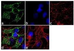
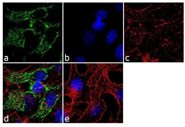
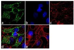
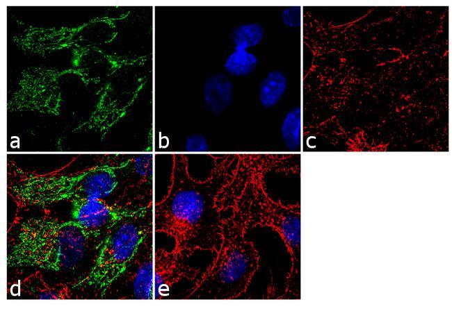
- Immunofluorescence analysis was performed on fixed and permeabilized HepG2 cells for detection of endogenous Glutaminase using Anti- Glutaminase Recombinant Rabbit Monoclonal Antibody (Product # 701965, 2 µg/mL) and labeled with Goat anti-Rabbit IgG (H+L) Superclonal™ Secondary Antibody, Alexa Fluor® 488 conjugate (Product # A27034, 1:2000). Panel a) shows representative cells that were stained for detection and localization of Glutaminase (green), Panel b) is stained for nuclei (blue) using SlowFade® Gold Antifade Mountant with DAPI (Product # S36938). Panel c) represents cytoskeletal F-actin staining using Alexa Fluor® 555 Rhodamine Phalloidin (Product # R415, 1:300). Panel d) is a composite image of Panels a, b and c clearly demonstrating cytoplasmic localization of Glutaminase. Panel e) represents control cells with no primary antibody to assess background. The images were captured at 60X magnification.
- Submitted by
- Invitrogen Antibodies (provider)
- Main image
- Experimental details
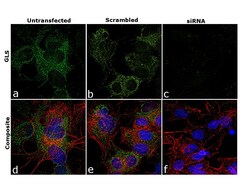
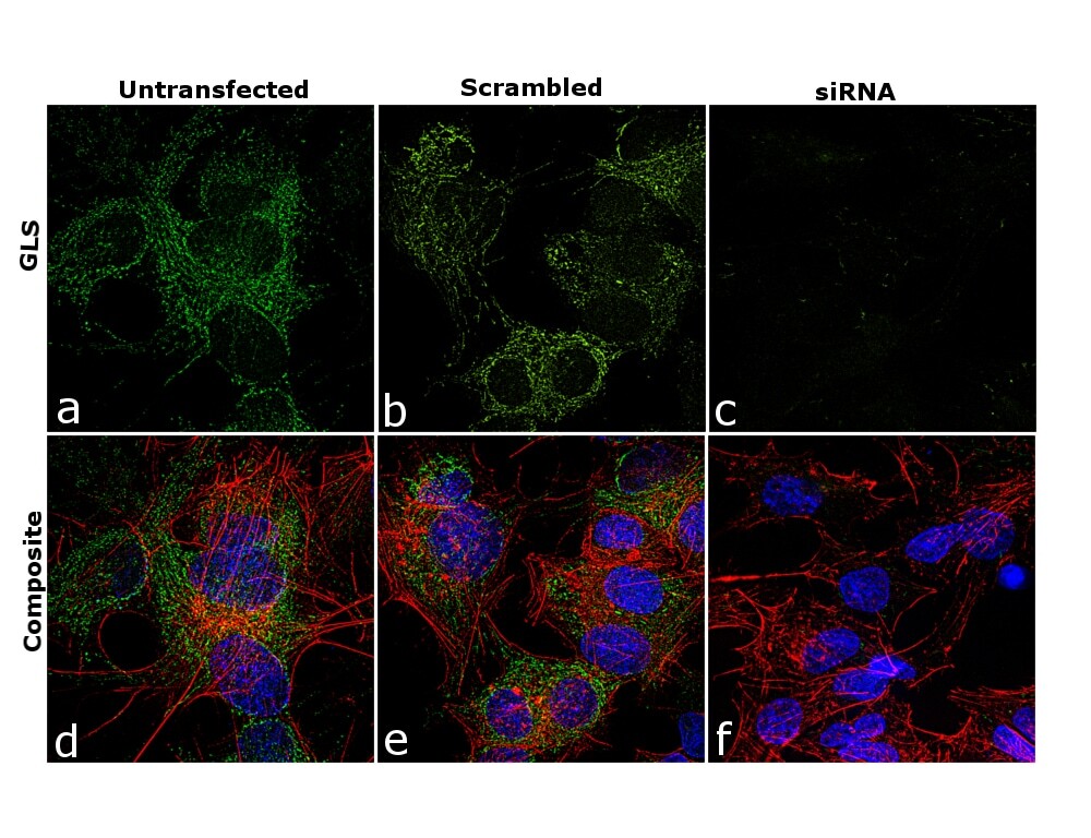
- Knockdown of GLS was achieved by transfecting Hep G2 cells with GLS specific siRNAs (Silencer® select Product # s5839, s5840). Immunofluorescence analysis was performed using untransfected Hep G2 cells (panels a, d), transfected with non-specific scrambled siRNA (panels b,e) and transfected with GLS specific siRNAs (panel c,f). Cells were fixed, permeabilized, and probed with GLS (6H5L15), Recombinant Rabbit Monoclonal Antibody (Product # 701965, 5 µg/mL), followed by labelling with Goat anti-Rabbit IgG (H+L) Superclonal™ Secondary Antibody, Alexa Fluor® 488 conjugate (Product # A27034; 1:2000 dilution) for 45 minutes at room temperature. Nuclei (blue) were stained using ProLong™ Diamond Antifade Mountant with DAPI (Product # P36962) and Rhodamine Phalloidin (Product # R415, 1:300) was used for cytoskeletal F-actin (red) staining. Reduction of mitochondrial GLS expression was observed upon siRNA mediated knockdown (panel c,f) confirming the specificity of the antibody to GLS. The images were captured at 60X magnification.

- Submitted by
- Invitrogen Antibodies (provider)
- Main image
- Experimental details
- Immunofluorescence analysis was performed on fixed and permeabilized HepG2 cells for detection of endogenous Glutaminase using Anti- Glutaminase Recombinant Rabbit Monoclonal Antibody (Product # 701965, 2 µg/mL) and labeled with Goat anti-Rabbit IgG (Heavy Chain) Superclonal™ Secondary Antibody, Alexa Fluor® 488 conjugate (Product # A27034, 1:2000). Panel a) shows representative cells that were stained for detection and localization of Glutaminase (green), Panel b) is stained for nuclei (blue) using SlowFade® Gold Antifade Mountant with DAPI (Product # S36938). Panel c) represents cytoskeletal F-actin staining using Alexa Fluor® 555 Rhodamine Phalloidin (Product # R415, 1:300). Panel d) is a composite image of Panels a, b and c clearly demonstrating cytoplasmic localization of Glutaminase. Panel e) represents control cells with no primary antibody to assess background. The images were captured at 60X magnification.
- Submitted by
- Invitrogen Antibodies (provider)
- Main image

- Experimental details
- Knockdown of GLS was achieved by transfecting Hep G2 cells with GLS specific siRNAs (Silencer® select Product # s5839, s5840). Immunofluorescence analysis was performed using untransfected Hep G2 cells (panels a, d), transfected with non-specific scrambled siRNA (panels b,e) and transfected with GLS specific siRNAs (panel c,f). Cells were fixed, permeabilized, and probed with GLS (6H5L15), Recombinant Rabbit Monoclonal Antibody (Product # 701965, 5 µg/mL), followed by labelling with Goat anti-Rabbit IgG (Heavy Chain) Superclonal™ Secondary Antibody, Alexa Fluor® 488 conjugate (Product # A27034; 1:2000 dilution) for 45 minutes at room temperature. Nuclei (blue) were stained using ProLong™ Diamond Antifade Mountant with DAPI (Product # P36962) and Rhodamine Phalloidin (Product # R415, 1:300) was used for cytoskeletal F-actin (red) staining. Reduction of mitochondrial GLS expression was observed upon siRNA mediated knockdown (panel c,f) confirming the specificity of the antibody to GLS. The images were captured at 60X magnification.
Supportive validation
- Submitted by
- Invitrogen Antibodies (provider)
- Main image
- Experimental details
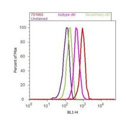
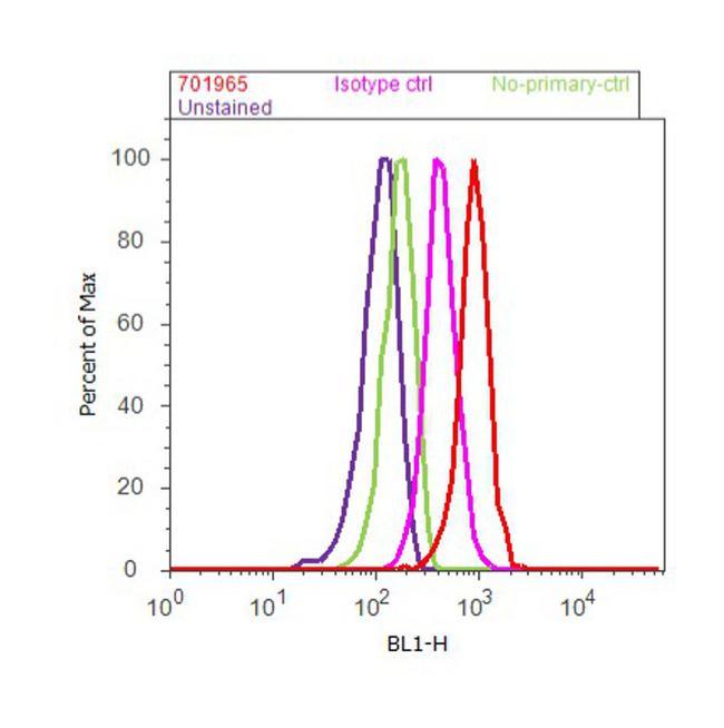
- Flow Cytometry analysis of endogenous Glutaminase was performed on HeLa cells labeled with ABfinity™ Anti-Glutaminase Recombinant Rabbit Monoclonal Antibody (Product# 701965, 5 ug/ 1M cells) or with rabbit isotype control at 0.5 ug/ml and detected with Goat anti-Rabbit IgG (H+L) Superclonal™ Secondary Antibody, (Alexa Fluor® 488 conjugate, Product# A27034, 0.4 ug/ml, 1:2500) as represented by the red and pink histograms respectively. The purple histogram represents unstained control cells and the green histogram represents no-primary-antibody control. A representative of 10,000 cells were acquired and analyzed for each sample using an Attune® Acoustic Focusing Cytometer (4468770).
Supportive validation
- Submitted by
- Invitrogen Antibodies (provider)
- Main image
- Experimental details
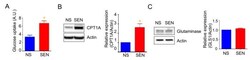
- Figure 2 Mitochondrial fuel usage is altered during doxorubicin-induced senescence. ( A ) Glucose uptake rate was measured by 2-NBDG, a fluorescently labeled deoxyglucose analog. Quantification and representative western blots of ( B ) carnitine palmitoyltransferase I (CPT1A) and ( C ) glutaminase (GLS1), both showing beta-actin as a loading control. Data are reported as mean +- SEM of three to six independent experiments. Significant differences were determined by Student's t -tests. *p
