Explore
 Validate
 Learn
Learn Western blot
 ELISA
ELISAAntibody data
- Antibody Data
- Antigen structure
- References [2]
- Comments [0]
- Validations
- Western blot [1]
Submit
Validation data
Reference
Comment
Report error
- Product number
- A01272-2 - Provider product page

- Provider
- Boster Biological Technology
- Product name
- Anti-Glutaminase/GLS Antibody Picoband™
- Antibody type
- Polyclonal
- Description
- Rabbit IgG polyclonal antibody for Glutaminase/GLS detection. Tested with WB, IHC-P, ICC/IF, FCM, Direct ELISA in Human;Mouse;Monkey;Rat.
- Reactivity
- Human, Mouse, Rat, Simian
- Host
- Rabbit
- Vial size
- 100μg/vial
- Concentration
- Add 0.2ml of distilled water will yield a concentration of 500ug/ml.
- Storage
- At -20°C for one year. After reconstitution, at 4°C for one month. It can also be aliquoted and stored frozen at -20°C for a longer time. Avoid repeated freezing and thawing.
- Handling
- Add 0.2ml of distilled water will yield a concentration of 500ug/ml.
Submitted references Microbiome and spatially resolved metabolomics analysis reveal the anticancer role of gut Akkermansia muciniphila by crosstalk with intratumoral microbiota and reprogramming tumoral metabolism in mice.
Safflower Yellow Improves the Synaptic Structural Plasticity by Ameliorating the Disorder of Glutamate Circulation in Aβ(1-42)-induced AD Model Rats.
Zhu Z, Cai J, Hou W, Xu K, Wu X, Song Y, Bai C, Mo YY, Zhang Z
Gut microbes 2023 Jan-Dec;15(1):2166700
Gut microbes 2023 Jan-Dec;15(1):2166700
Safflower Yellow Improves the Synaptic Structural Plasticity by Ameliorating the Disorder of Glutamate Circulation in Aβ(1-42)-induced AD Model Rats.
Hou J, Wang C, Zhang M, Ren M, Yang G, Qu Z, Hu Y
Neurochemical research 2020 Aug;45(8):1870-1887
Neurochemical research 2020 Aug;45(8):1870-1887
No comments: Submit comment
Supportive validation
- Submitted by
- Boster Biological Technology (provider)
- Main image

- Experimental details
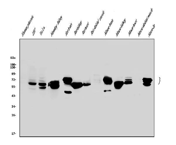
- Western blot analysis of Glutaminase/GLS using anti-Glutaminase/GLS antibody (A01272-2). Electrophoresis was performed on a 5-20% SDS-PAGE gel at 70V (Stacking gel) / 90V (Resolving gel) for 2-3 hours. The sample well of each lane was loaded with 50ug of sample under reducing conditions. Lane 1: human placenta tissue lysates, Lane 2: human U87 whole cell lysates, Lane 3: human Hela whole cell lysates, Lane 4: monkey kidney tissue lysates, Lane 5: rat brain tissue lysates, Lane 6: rat kidney tissue lysates, Lane 7: rat heart tissue lysates. Lane 8: rat skeletal muscle tissue lysates. Lane 9: mouse brain tissue lysates. Lane 10: mouse kidney tissue lysates. Lane 11: mouse heart tissue lysates. Lane 12: mouse skeletal muscle tissue lysates. Lane 13: mouse Neuro-2a whole cell lysates. After Electrophoresis, proteins were transferred to a Nitrocellulose membrane at 150mA for 50-90 minutes. Blocked the membrane with 5% Non-fat Milk/ TBS for 1.5 hour at RT. The membrane was incubated with rabbit anti-Glutaminase/GLS antigen affinity purified polyclonal antibody (Catalog # A01272-2) at 0.25 μg/mL overnight at 4°C, then washed with TBS-0.1%Tween 3 times with 5 minutes each and probed with a goat anti-rabbit IgG-HRP secondary antibody at a dilution of 1:10000 for 1.5 hour at RT. The signal is developed using an Enhanced Chemiluminescent detection (ECL) kit (Catalog # EK1002) with Tanon 5200 system. A specific band was detected for Glutaminase/GLS at approximately 65-73KD. The expected band size for Glutaminase/GLS is at 73KD.
- Additional image