Explore
 Validate
 Learn
Learn Western blot
 Immunocytochemistry
Antibody data
- Antibody Data
- Antigen structure
- References [2]
- Comments [0]
- Validations
- Immunocytochemistry [1]
- Immunohistochemistry [4]
- Other assay [1]
Submit
Validation data
Reference
Comment
Report error
- Product number
- PA5-29663 - Provider product page

- Provider
- Invitrogen Antibodies
- Product name
- GATA4 Polyclonal Antibody
- Antibody type
- Polyclonal
- Antigen
- Recombinant full-length protein
- Description
- Recommended positive controls: HepG2 nuclear extract, mouse heart. Predicted reactivity: Mouse (89%), Rat (89%), Xenopus laevis (82%), Dog (96%), Pig (93%), Chicken (85%), Bovine (95%). Store product as a concentrated solution. Centrifuge briefly prior to opening the vial.
- Reactivity
- Human, Mouse
- Host
- Rabbit
- Isotype
- IgG
- Vial size
- 100 μL
- Concentration
- 0.21 mg/mL
- Storage
- Store at 4°C short term. For long term storage, store at -20°C, avoiding freeze/thaw cycles.
Submitted references Epigenetic Changes Governing Scn5a Expression in Denervated Skeletal Muscle.
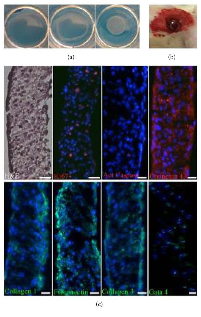
C-Kit Cardiac Progenitor Cell Based Cell Sheet Improves Vascularization and Attenuates Cardiac Remodeling following Myocardial Infarction in Rats.
Carreras D, Martinez-Moreno R, Pinsach-Abuin ML, Santafe MM, Gomà P, Brugada R, Scornik FS, Pérez GJ, Pagans S
International journal of molecular sciences 2021 Mar 9;22(5)
International journal of molecular sciences 2021 Mar 9;22(5)
C-Kit Cardiac Progenitor Cell Based Cell Sheet Improves Vascularization and Attenuates Cardiac Remodeling following Myocardial Infarction in Rats.
Dergilev K, Tsokolaeva Z, Makarevich P, Beloglazova I, Zubkova E, Boldyreva M, Ratner E, Dyikanov D, Menshikov M, Ovchinnikov A, Ageev F, Parfyonova Y
BioMed research international 2018;2018:3536854
BioMed research international 2018;2018:3536854
No comments: Submit comment
Supportive validation
- Submitted by
- Invitrogen Antibodies (provider)
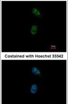
- Main image

- Experimental details
- Immunofluorescent analysis of GATA4 in paraformaldehyde-fixed HeLa cells using a GATA4 polyclonal antibody (Product # PA5-29663) at a 1:500 dilution.
Supportive validation
- Submitted by
- Invitrogen Antibodies (provider)
- Main image
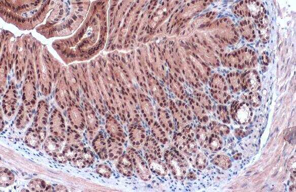
- Experimental details
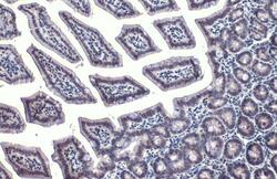
- Immunohistochemistry (Paraffin) analysis of GATA4 was performed in paraffin-embedded mouse duodenum tissue using GATA4 Polyclonal Antibody (Product # PA5-29663) at a dilution of 1:1000. Antigen Retrieval: Citrate buffer, pH 6.0, 15 min.
- Submitted by
- Invitrogen Antibodies (provider)
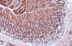
- Main image
- Experimental details
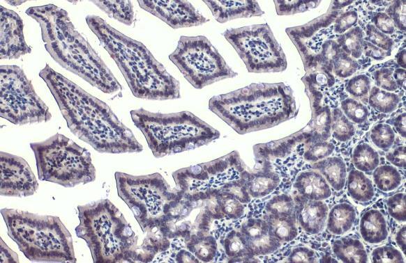
- GATA4 Polyclonal Antibody detects GATA4 protein at nucleus by immunohistochemical analysis. Sample: Paraffin-embedded mouse colon. GATA4 stained by GATA4 Polyclonal Antibody (Product # PA5-29663) diluted at 1:500. Antigen Retrieval: Citrate buffer, pH 6.0, 15 min.
- Submitted by
- Invitrogen Antibodies (provider)
- Main image

- Experimental details
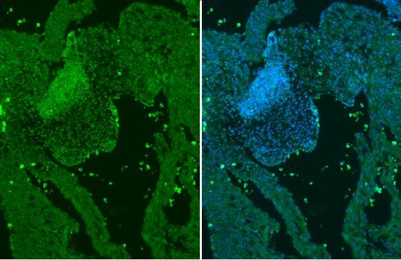
- GATA4 Polyclonal Antibody detects GATA4 protein at nucleus by immunohistochemical analysis. Sample: Paraffin-embedded mouse E13.5 embryo. Green: GATA4 stained by GATA4 Polyclonal Antibody (Product # PA5-29663) diluted at 1:250. Blue: Fluoroshield with DAPI. Antigen Retrieval: Citrate buffer, pH 6.0, 15 min.
- Submitted by
- Invitrogen Antibodies (provider)
- Main image
- Experimental details
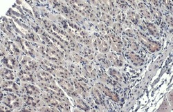
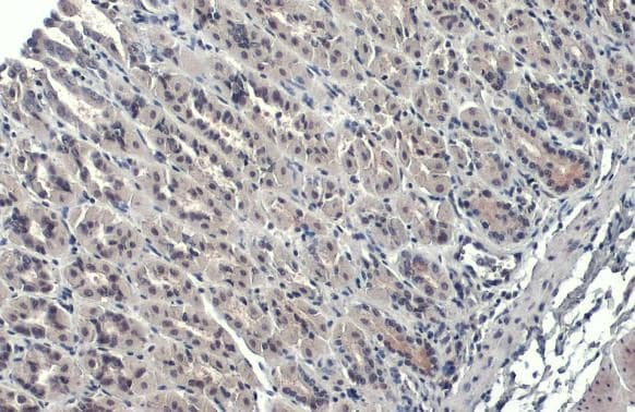
- GATA4 Polyclonal Antibody detects GATA4 protein at nucleus by immunohistochemical analysis. Sample: Paraffin-embedded mouse stomach. GATA4 stained by GATA4 Polyclonal Antibody (Product # PA5-29663) diluted at 1:500. Antigen Retrieval: Citrate buffer, pH 6.0, 15 min.
Supportive validation
- Submitted by
- Invitrogen Antibodies (provider)
- Main image

- Experimental details
- Figure 2 Isolation and characterization of cell sheets from c-kit+ CPC. (a) CPC sheets formed after 3 days of culture detached at room temperature onto thermoresponsive surfaces yielding a scaffold-free monolayered cell sheet. (b) Hematoxylin-eosin staining shows that the cell sheet represents a well-organized structure. CPC in the cell sheets survived, retained the ability to proliferate (Ki67), expressed progenitor cell marker Gata 4, interconnected with each other via connexin-43 gap junctions, and were surrounded extracellular matrix proteins (collagen 1, collagen 3, and fibronectin). (c) Macroscopic visualization of epicardially attached CPC sheet after implantation.