Explore
 Validate
 Learn
Learn Western blot
Antibody data
- Antibody Data
- Antigen structure
- References [6]
- Comments [0]
- Validations
- Western blot [2]
- Immunocytochemistry [10]
- Immunohistochemistry [3]
- Flow cytometry [5]
- Other assay [3]
Submit
Validation data
Reference
Comment
Report error
- Product number
- PA1-102X - Provider product page

- Provider
- Invitrogen Antibodies
- Product name
- GATA4 Polyclonal Antibody
- Antibody type
- Polyclonal
- Antigen
- Synthetic peptide
- Description
- PA1-102 has been successfully used in Western blot, FACS, immunohistochemistry (paraffin) and immunofluorescence applications with human and mouse samples. PA1-102 can be used for immunofluorescence analysis of GATA4 in the endoderm derived from human iPSCs.
- Reactivity
- Human, Mouse
- Host
- Rabbit
- Isotype
- IgG
- Vial size
- 20 μL
- Concentration
- 1 mg/mL
- Storage
- -20°C
Submitted references Neuregulin-1, in a Conducive Milieu with Wnt/BMP/Retinoic Acid, Prolongs the Epicardial-Mediated Cardiac Regeneration Capacity of Neonatal Heart Explants.
Androgen Receptor, Although Not a Specific Marker For, Is a Novel Target to Suppress Glioma Stem Cells as a Therapeutic Strategy for Glioblastoma.
Scalable Biomimetic Coaxial Aligned Nanofiber Cardiac Patch: A Potential Model for "Clinical Trials in a Dish".
Human Cardiac Progenitor Cells Enhance Exosome Release and Promote Angiogenesis Under Physoxia.
Stoichiometric optimization of Gata4, Hand2, Mef2c, and Tbx5 expression for contractile cardiomyocyte reprogramming.
Generation and characterization of a human iPSC line from an ALS patient carrying the Q66K-MATR3 mutation.
Arora H, Lavin AC, Balkan W, Hare JM, White IA
Journal of stem cells & regenerative medicine 2021;17(1):18-27
Journal of stem cells & regenerative medicine 2021;17(1):18-27
Androgen Receptor, Although Not a Specific Marker For, Is a Novel Target to Suppress Glioma Stem Cells as a Therapeutic Strategy for Glioblastoma.
Zhao N, Wang F, Ahmed S, Liu K, Zhang C, Cathcart SJ, DiMaio DJ, Punsoni M, Guan B, Zhou P, Wang S, Batra SK, Bronich T, Hei TK, Lin C, Zhang C
Frontiers in oncology 2021;11:616625
Frontiers in oncology 2021;11:616625
Scalable Biomimetic Coaxial Aligned Nanofiber Cardiac Patch: A Potential Model for "Clinical Trials in a Dish".
Kumar N, Sridharan D, Palaniappan A, Dougherty JA, Czirok A, Isai DG, Mergaye M, Angelos MG, Powell HM, Khan M
Frontiers in bioengineering and biotechnology 2020;8:567842
Frontiers in bioengineering and biotechnology 2020;8:567842
Human Cardiac Progenitor Cells Enhance Exosome Release and Promote Angiogenesis Under Physoxia.
Dougherty JA, Patel N, Kumar N, Rao SG, Angelos MG, Singh H, Cai C, Khan M
Frontiers in cell and developmental biology 2020;8:130
Frontiers in cell and developmental biology 2020;8:130
Stoichiometric optimization of Gata4, Hand2, Mef2c, and Tbx5 expression for contractile cardiomyocyte reprogramming.
Zhang Z, Zhang W, Nam YJ
Scientific reports 2019 Oct 18;9(1):14970
Scientific reports 2019 Oct 18;9(1):14970
Generation and characterization of a human iPSC line from an ALS patient carrying the Q66K-MATR3 mutation.
Pollini D, Loffredo R, Cardano M, Conti L, Lattante S, Notarangelo A, Sabatelli M, Provenzani A
Stem cell research 2018 Dec;33:146-150
Stem cell research 2018 Dec;33:146-150
No comments: Submit comment
Supportive validation
- Submitted by
- Invitrogen Antibodies (provider)
- Main image

- Experimental details
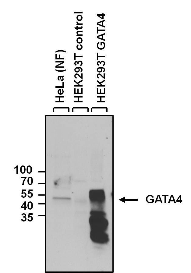
- Western blot analysis of GATA4 was performed by loading 40 µg of HeLa nuclear fraction (NF) (Lane 1), HEK293T control (Lane 2), and HEK293T GATA4 over-expression lysate (Lane 3) onto a 4-20% Tris-HCl polyacrylamide gel. Proteins were transferred to a PVDF membrane and blocked with 5% BSA/TBST for 1 hour. Membranes were probed with a rabbit polyclonal antibody recognizing GATA4 (Product # PA1-102) at a dilution of 1:1000 overnight at 4°C on a rocking platform. Membranes were washed in TBS-0.1%Tween 20 and probed with a goat anti-rabbit-HRP secondary antibody (Product # 31460) at a dilution of 1:20,000 for one hour. Membranes were washed and chemiluminescent detection performed using Super Signal West Pico (Product # 34078). Nuclear fractions were generated using the NE-PER kit (Product # 78833).
- Submitted by
- Invitrogen Antibodies (provider)
- Main image
- Experimental details
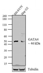
- Western blot analysis was performed on whole cell extracts (20 µg lysate) of SH-SY5Y (Lane 1) and Hep G2 (Lane 2). The blots were probed with Anti-GATA4 Rabbit Polyclonal Antibody (Product # PA1-102, 1:500-1:2000 dilution) and detected by chemiluminescence using Goat anti-Rabbit IgG (Heavy Chain) Superclonal™ Secondary Antibody, HRP conjugate (Product # A27036, 0.4 µg/mL, 1:2500 dilution). A 46 kDa band corresponding to GATA4 was observed across the cell lines tested. Known quantity of protein samples were electrophoresed using Novex® NuPAGE® 12 % Bis-Tris gel (Product # NP0342BOX), XCell SureLock™ Electrophoresis System (Product # EI0002) and Novex® Sharp Pre-Stained Protein Standard (Product # LC5800). Resolved proteins were then transferred onto a nitrocellulose membrane with iBlot® 2 Dry Blotting System (Product # IB21001). The membrane was probed with the relevant primary and secondary Antibody following blocking with 5 % skimmed milk. Chemiluminescent detection was performed using Pierce™ ECL Western Blotting Substrate (Product # 32106).

Supportive validation
- Submitted by
- Invitrogen Antibodies (provider)
- Main image
- Experimental details
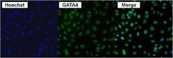
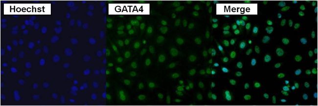
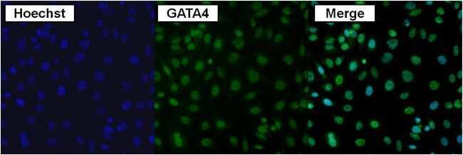
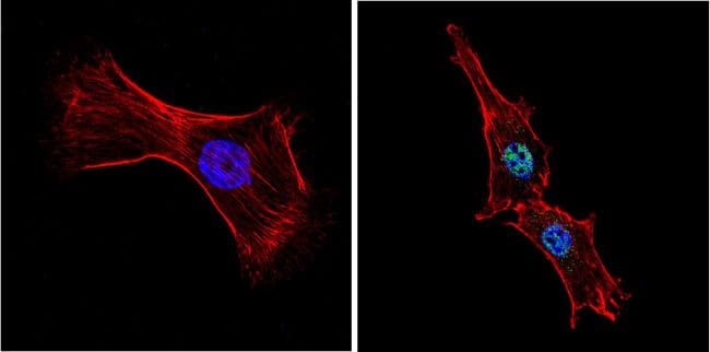
- Immunofluorescent analysis of GATA4 using anti-GATA4 polyclonal antibody (Product # PA1-102) in HeLa cells. Formalin fixed cells were permeabilized with 0.1% Triton X-100 in TBS for 10 minutes at room temperature. Cells were blocked with Blocker BSA (Product # 37525) for 15 minutes at room temperature and probed with a rabbit polyclonal antibody recognizing GATA4 (Product # PA1-102), at a dilution of 1:50 for 1 hour at room temperature. Cells were then washed with PBS and incubated with DyLight 488 (green) goat-anti-rabbit secondary antibody (Product # 35552) at a dilution of 1:400 for 30 minutes at room temperature. Nuclei (blue) were stained with Hoechst 33342 dye (Product # 62249). Images were taken on a Thermo Scientific ArrayScan at 20X magnification.
- Submitted by
- Invitrogen Antibodies (provider)
- Main image
- Experimental details
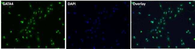
- Immunofluorescent analysis of GATA4 (green) in embryoid body endoderm generated from Gibco ® Human Episomal iPSC Line grown on Geltrex® in Essential 8TM Medium. After 2 weeks in culture, EB were dissociated with TrypLETM and re-plated onto Geltrex®-coated multi-well plates. Cells were fixed, permeabilized and blocked for immunostaining. Cells were stained with a GATA4 polyclonal antibody (Product # PA1-102) at a dilution of 1:100 in 3% BSA/PBS blocking buffer overnight at 4°C, and then incubated with Alexa Fluor® 488 donkey anti rabbit antibody (Product # A-21206) at a 1:500 dilution in conjunction with NucBlue® Fixed Cell Ready Probes® Reagent. After another 3 washes, images were taken on EVOS®Floid® Cell Imaging system at 10X magnification.
- Submitted by
- Invitrogen Antibodies (provider)
- Main image
- Experimental details
- Immunofluorescent analysis of GATA4 using anti-GATA4 polyclonal antibody (Product # PA1-102) in HeLa cells. Formalin fixed cells were permeabilized with 0.1% Triton X-100 in TBS for 10 minutes at room temperature. Cells were blocked with Blocker BSA (Product # 37525) for 15 minutes at room temperature and probed with a rabbit polyclonal antibody recognizing GATA4 (Product # PA1-102), at a dilution of 1:50 for 1 hour at room temperature. Cells were then washed with PBS and incubated with DyLight 488 (green) goat-anti-rabbit secondary antibody (Product # 35552) at a dilution of 1:400 for 30 minutes at room temperature. Nuclei (blue) were stained with Hoechst 33342 dye (Product # 62249). Images were taken on a Thermo Scientific ArrayScan at 20X magnification.
- Submitted by
- Invitrogen Antibodies (provider)
- Main image

- Experimental details
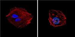
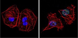
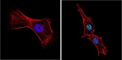
- Immunofluorescent analysis of GATA4 (green) showing positive staining in the nucleus of Hela cells (right) compared with a negative control in the absence of primary antibody (left). Formalin-fixed cells were permeabilized with 0.1% Triton X-100 in TBS for 5-10 minutes, blocked with 3% BSA-PBS for 30 minutes at room temperature and probed with a GATA4 polyclonal antibody (Product # PA1-102) in 3% BSA-PBS at a dilution of 1:200 and incubated overnight at 4 ºC in a humidified chamber. Cells were washed with PBST and incubated with a DyLight 488-conjugated goat-anti-rabbit IgG secondary antibody in PBS at room temperature in the dark. F-actin (red) was stained with a fluorescent red phalloidin and nuclei (blue) were stained with DAPI for 5-10 minutes in the dark. Images were taken at a magnification of 60x.

- Submitted by
- Invitrogen Antibodies (provider)
- Main image
- Experimental details
- Immunofluorescent analysis of GATA4 (green) in embryoid body endoderm generated from Gibco ® Human Episomal iPSC Line grown on Geltrex® in Essential 8TM Medium. After 2 weeks in culture, EB were dissociated with TrypLETM and re-plated onto Geltrex®-coated multi-well plates. Cells were fixed, permeabilized and blocked for immunostaining. Cells were stained with a GATA4 polyclonal antibody (Product # PA1-102) at a dilution of 1:100 in 3% BSA/PBS blocking buffer overnight at 4°C, and then incubated with Alexa Fluor® 488 donkey anti rabbit antibody (Product # A-21206) at a 1:500 dilution in conjunction with NucBlue® Fixed Cell Ready Probes® Reagent. After another 3 washes, images were taken on EVOS®Floid® Cell Imaging system at 10X magnification.

- Submitted by
- Invitrogen Antibodies (provider)
- Main image
- Experimental details
- Immunofluorescent analysis of GATA4 (green) showing positive staining in the nucleus of HepG2 cells (right) compared with a negative control in the absence of primary antibody (left). Formalin-fixed cells were permeabilized with 0.1% Triton X-100 in TBS for 5-10 minutes, blocked with 3% BSA-PBS for 30 minutes at room temperature and probed with a GATA4 polyclonal antibody (Product # PA1-102) in 3% BSA-PBS at a dilution of 1:200 and incubated overnight at 4 ºC in a humidified chamber. Cells were washed with PBST and incubated with a DyLight 488-conjugated goat-anti-rabbit IgG secondary antibody in PBS at room temperature in the dark. F-actin (red) was stained with a fluorescent red phalloidin and nuclei (blue) were stained with DAPI for 5-10 minutes in the dark. Images were taken at a magnification of 60x.

- Submitted by
- Invitrogen Antibodies (provider)
- Main image
- Experimental details
- Immunofluorescent analysis of GATA4 (green) showing positive staining in the nucleus of Hela cells (right) compared with a negative control in the absence of primary antibody (left). Formalin-fixed cells were permeabilized with 0.1% Triton X-100 in TBS for 5-10 minutes, blocked with 3% BSA-PBS for 30 minutes at room temperature and probed with a GATA4 polyclonal antibody (Product # PA1-102) in 3% BSA-PBS at a dilution of 1:200 and incubated overnight at 4 ºC in a humidified chamber. Cells were washed with PBST and incubated with a DyLight 488-conjugated goat-anti-rabbit IgG secondary antibody in PBS at room temperature in the dark. F-actin (red) was stained with a fluorescent red phalloidin and nuclei (blue) were stained with DAPI for 5-10 minutes in the dark. Images were taken at a magnification of 60x.

- Submitted by
- Invitrogen Antibodies (provider)
- Main image

- Experimental details
- Immunofluorescent analysis of GATA4 (green) showing positive staining in the nucleus of HepG2 cells (right) compared with a negative control in the absence of primary antibody (left). Formalin-fixed cells were permeabilized with 0.1% Triton X-100 in TBS for 5-10 minutes, blocked with 3% BSA-PBS for 30 minutes at room temperature and probed with a GATA4 polyclonal antibody (Product # PA1-102) in 3% BSA-PBS at a dilution of 1:200 and incubated overnight at 4 ºC in a humidified chamber. Cells were washed with PBST and incubated with a DyLight 488-conjugated goat-anti-rabbit IgG secondary antibody in PBS at room temperature in the dark. F-actin (red) was stained with a fluorescent red phalloidin and nuclei (blue) were stained with DAPI for 5-10 minutes in the dark. Images were taken at a magnification of 60x.

- Submitted by
- Invitrogen Antibodies (provider)
- Main image
- Experimental details
- Immunofluorescent analysis of GATA4 (green) showing positive staining in the nucleus of NIH-3T3 cells (right) compared with a negative control in the absence of primary antibody (left). Formalin-fixed cells were permeabilized with 0.1% Triton X-100 in TBS for 5-10 minutes, blocked with 3% BSA-PBS for 30 minutes at room temperature and probed with a GATA4 polyclonal antibody (Product # PA1-102) in 3% BSA-PBS at a dilution of 1:100 and incubated overnight at 4 ºC in a humidified chamber. Cells were washed with PBST and incubated with a DyLight 488-conjugated goat-anti-rabbit IgG secondary antibody in PBS at room temperature in the dark. F-actin (red) was stained with a fluorescent red phalloidin and nuclei (blue) were stained with DAPI for 5-10 minutes in the dark. Images were taken at a magnification of 60x.
- Submitted by
- Invitrogen Antibodies (provider)
- Main image

- Experimental details
- Immunofluorescent analysis of GATA4 (green) in embryoid body endoderm generated from Gibco ® Human Episomal iPSC Line grown on Geltrex® in Essential 8TM Medium. After 2 weeks in culture, EB were dissociated with TrypLETM and re-plated onto Geltrex®-coated multi-well plates. Cells were fixed, permeabilized and blocked for immunostaining. Cells were stained with a GATA4 polyclonal antibody (Product # PA1-102) at a dilution of 1:100 in 3% BSA/PBS blocking buffer overnight at 4°C, and then incubated with Alexa Fluor® 488 donkey anti rabbit antibody (Product # A-21206) at a 1:500 dilution in conjunction with NucBlue® Fixed Cell Ready Probes® Reagent. After another 3 washes, images were taken on EVOS®Floid® Cell Imaging system at 10X magnification.

Supportive validation
- Submitted by
- Invitrogen Antibodies (provider)
- Main image
- Experimental details
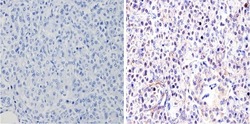
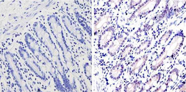
- Immunohistochemistry analysis of GATA4 showing positive staining in the nucleus of paraffin-treated Human pancreas tissue (right) compared with a negative control in the absence of primary antibody (left). To expose target proteins, antigen retrieval method was performed using 10mM sodium citrate (pH 6.0) microwaved for 8-15 min. Following antigen retrieval, tissues were blocked in 3% H2O2-methanol for 15 min at room temperature, washed with ddH2O and PBS, and then probed with a GATA4 polyclonal antibody (Product # PA1-102) diluted by 3% BSA-PBS at a dilution of 1:200 overnight at 4°C in a humidified chamber. Tissues were washed extensively PBST and detection was performed using an HRP-conjugated secondary antibody followed by colorimetric detection using a DAB kit. Tissues were counterstained with hematoxylin and dehydrated with ethanol and xylene to prep for mounting.
- Submitted by
- Invitrogen Antibodies (provider)
- Main image
- Experimental details
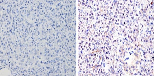
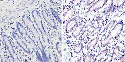
- Immunohistochemistry analysis of GATA4 showing positive staining in the nucleus of paraffin-treated Human small intestine (right) compared with a negative control in the absence of primary antibody (left). To expose target proteins, antigen retrieval method was performed using 10mM sodium citrate (pH 6.0) microwaved for 8-15 min. Following antigen retrieval, tissues were blocked in 3% H2O2-methanol for 15 min at room temperature, washed with ddH2O and PBS, and then probed with a GATA4 polyclonal antibody (Product # PA1-102) diluted by 3% BSA-PBS at a dilution of 1:200 overnight at 4°C in a humidified chamber. Tissues were washed extensively PBST and detection was performed using an HRP-conjugated secondary antibody followed by colorimetric detection using a DAB kit. Tissues were counterstained with hematoxylin and dehydrated with ethanol and xylene to prep for mounting.
- Submitted by
- Invitrogen Antibodies (provider)
- Main image
- Experimental details
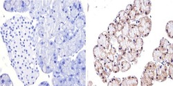
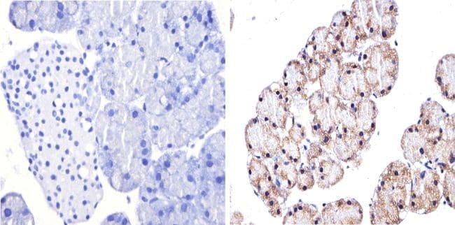
- Immunohistochemistry analysis of GATA4 showing positive staining in the nucleus of paraffin-treated Mouse pancreas tissue (right) compared with a negative control in the absence of primary antibody (left). To expose target proteins, antigen retrieval method was performed using 10mM sodium citrate (pH 6.0) microwaved for 8-15 min. Following antigen retrieval, tissues were blocked in 3% H2O2-methanol for 15 min at room temperature, washed with ddH2O and PBS, and then probed with a GATA4 polyclonal antibody (Product # PA1-102) diluted by 3% BSA-PBS at a dilution of 1:500 overnight at 4°C in a humidified chamber. Tissues were washed extensively PBST and detection was performed using an HRP-conjugated secondary antibody followed by colorimetric detection using a DAB kit. Tissues were counterstained with hematoxylin and dehydrated with ethanol and xylene to prep for mounting.
Supportive validation
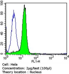
- Submitted by
- Invitrogen Antibodies (provider)
- Main image
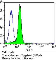
- Experimental details
- Flow cytometry analysis of GATA4 in Hela cells compared to an isotype control (blue). Cells were harvested, adjusted to a concentration of 1-5x10^6 cells/mL, fixed with 2% paraformaldehyde and washed with PBS. Cells were penetrated by dropping the supernatant, adding 90% methanol and incubated for 10 minutes at room temperature. Follwing penetration, cells were blocked with a 2% solution of BSA-PBS for 30 min at room temperature and incubated with a GATA4 polyclonal antibody (Product # PA1-102) at a dilution of 2 µg/test for 60 min at room temperature. Cells were then incubated for 40 min at room temperature in the dark using a Dylight 488-conjugated goat anti-rabbit IgG (H+L) secondary antibody and re-suspended in PBS for FACS analysis.
- Submitted by
- Invitrogen Antibodies (provider)
- Main image

- Experimental details
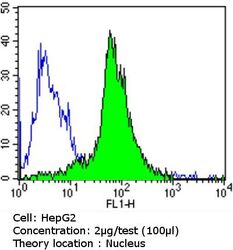
- Flow cytometry analysis of GATA4 in HepG2 cells compared to an isotype control (blue). Cells were harvested, adjusted to a concentration of 1-5x10^6 cells/mL, fixed with 2% paraformaldehyde and washed with PBS. Cells were penetrated by dropping the supernatant, adding 90% methanol and incubated for 10 minutes at room temperature. Follwing penetration, cells were blocked with a 2% solution of BSA-PBS for 30 min at room temperature and incubated with a GATA4 polyclonal antibody (Product # PA1-102) at a dilution of 2 µg/test for 60 min at room temperature. Cells were then incubated for 40 min at room temperature in the dark using a Dylight 488-conjugated goat anti-rabbit IgG (H+L) secondary antibody and re-suspended in PBS for FACS analysis.
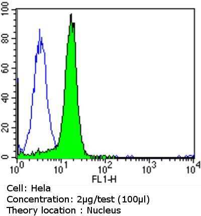
- Submitted by
- Invitrogen Antibodies (provider)
- Main image
- Experimental details
- Flow cytometry analysis of GATA4 in Hela cells compared to an isotype control (blue). Cells were harvested, adjusted to a concentration of 1-5x10^6 cells/mL, fixed with 2% paraformaldehyde and washed with PBS. Cells were penetrated by dropping the supernatant, adding 90% methanol and incubated for 10 minutes at room temperature. Follwing penetration, cells were blocked with a 2% solution of BSA-PBS for 30 min at room temperature and incubated with a GATA4 polyclonal antibody (Product # PA1-102) at a dilution of 2 µg/test for 60 min at room temperature. Cells were then incubated for 40 min at room temperature in the dark using a Dylight 488-conjugated goat anti-rabbit IgG (H+L) secondary antibody and re-suspended in PBS for FACS analysis.

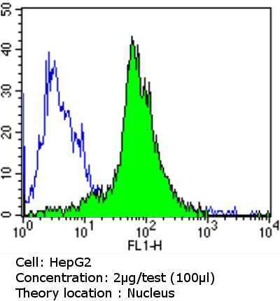
- Submitted by
- Invitrogen Antibodies (provider)
- Main image
- Experimental details
- Flow cytometry analysis of GATA4 in HepG2 cells compared to an isotype control (blue). Cells were harvested, adjusted to a concentration of 1-5x10^6 cells/mL, fixed with 2% paraformaldehyde and washed with PBS. Cells were penetrated by dropping the supernatant, adding 90% methanol and incubated for 10 minutes at room temperature. Follwing penetration, cells were blocked with a 2% solution of BSA-PBS for 30 min at room temperature and incubated with a GATA4 polyclonal antibody (Product # PA1-102) at a dilution of 2 µg/test for 60 min at room temperature. Cells were then incubated for 40 min at room temperature in the dark using a Dylight 488-conjugated goat anti-rabbit IgG (H+L) secondary antibody and re-suspended in PBS for FACS analysis.

- Submitted by
- Invitrogen Antibodies (provider)
- Main image
- Experimental details
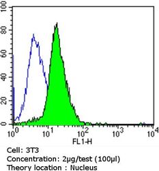
- Flow cytometry analysis of GATA4 in NIH/3T3 cells compared to an isotype control (blue). Cells were harvested, adjusted to a concentration of 1-5x10^6 cells/mL, fixed with 2% paraformaldehyde and washed with PBS. Cells were penetrated by dropping the supernatant, adding 90% methanol and incubated for 10 minutes at room temperature. Follwing penetration, cells were blocked with a 2% solution of BSA-PBS for 30 min at room temperature and incubated with a GATA4 polyclonal antibody (Product # PA1-102) at a dilution of 2 µg/test for 60 min at room temperature. Cells were then incubated for 40 min at room temperature in the dark using a Dylight 488-conjugated goat anti-rabbit IgG (H+L) secondary antibody and re-suspended in PBS for FACS analysis.

Supportive validation
- Submitted by
- Invitrogen Antibodies (provider)
- Main image
- Experimental details
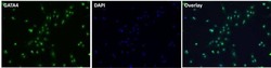
- NULL
- Submitted by
- Invitrogen Antibodies (provider)
- Main image
- Experimental details
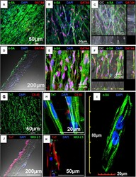
- FIGURE 4 Confocal imaging to assess the expression of cardiac markers in human induced-pluripotent stem cell-derived cardiomyocytes (hiPSC-CMs) cultured on the aligned coaxial nanofiber patch. (A-F) Confocal images showing the expression of alpha-SA and GATA4 in hiPSC-CMs cultured on aligned coaxial patches. (G,H) Confocal images showing the expression of TnT and Cx-43 in hiPSC-CMs cultured on aligned coaxial patches. (J,K) Confocal images showing the expression of TnT and Nkx2.5 in hiPSC-CMs cultured on aligned coaxial patches. (C,F) Z-stack images showing the distribution of the hiPSC-CMs through the depth of the coaxial patches. (D,J) Cross-section images of the coaxial patches showing the distribution of hiPSC-CMs in the patch. (I) Confocal image of a single hiPSC-CM cultured on an aligned coaxial patch.
- Submitted by
- Invitrogen Antibodies (provider)
- Main image
- Experimental details
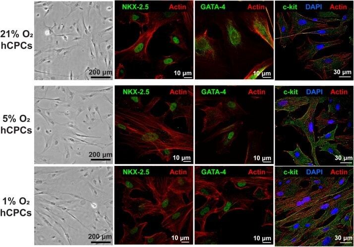
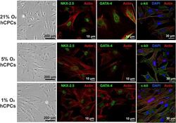
- FIGURE 1 hCPCs morphology and cardiac gene expression under normoxic and hypoxic microenvironments. Human CPCs were cultured under 21, 5, and 1% O 2 for 48 h. DIC imaging shows the typical morphology of cells, which is unchanged under hypoxia. Immunofluorescent staining for cardiac lineage markers NKX-2.5 (green, nuclear), GATA-4 (green, nuclear), and c-kit (green) showed their expression was maintained under all oxygen conditions. Nuclei are stained blue and F-actin is stained red.