Explore
 Validate
 Learn
Learn Western blot
Antibody data
- Antibody Data
- Antigen structure
- References [1]
- Comments [0]
- Validations
- Western blot [4]
- Immunocytochemistry [2]
- Immunohistochemistry [2]
- Other assay [1]
Submit
Validation data
Reference
Comment
Report error
- Product number
- PA5-86143 - Provider product page

- Provider
- Invitrogen Antibodies
- Product name
- Filamin A Polyclonal Antibody
- Antibody type
- Polyclonal
- Antigen
- Synthetic peptide
- Description
- Purity is > 95% by SDS-PAGE.
- Reactivity
- Human, Mouse, Rat
- Host
- Rabbit
- Isotype
- IgG
- Vial size
- 100 μL
- Concentration
- 1 mg/mL
- Storage
- Store at 4°C short term. For long term storage, store at -20°C, avoiding freeze/thaw cycles.
Submitted references Lipid-driven CFTR clustering is impaired in cystic fibrosis and restored by corrector drugs.
Abu-Arish A, Pandžić E, Luo Y, Sato Y, Turner MJ, Wiseman PW, Hanrahan JW
Journal of cell science 2022 Mar 1;135(5)
Journal of cell science 2022 Mar 1;135(5)
No comments: Submit comment
Supportive validation
- Submitted by
- Invitrogen Antibodies (provider)
- Main image

- Experimental details
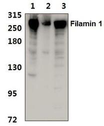
- Western blot analysis of Filamin A in Lane 1: HEK293T whole cell lysate, Lane 2: Raw264.7 whole cell lysate, Lane 3: H9C2 whole cell lysate. Sample was incubated with Filamin A polyclonal antibody (Product # PA5-86143) using a 1:500 dilution.
- Submitted by
- Invitrogen Antibodies (provider)
- Main image
- Experimental details
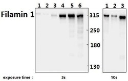
- Western blot analysis of Filamin A in Lane 1: mouse heart tissue (40 µg), Lane 2: rat heart tissue (40 µg), Lane 3: HEK293T whole cell lysate (40 µg), Lane 4: A549 whole cell lysate (40 µg), Lane 5: HCC827 whole cell lysate (40 µg), Lane 6: U87MG whole cell lysate (40 µg). Sample was incubated with Filamin A polyclonal antibody (Product # PA5-86143) using a 1:500 dilution.
- Submitted by
- Invitrogen Antibodies (provider)
- Main image
- Experimental details
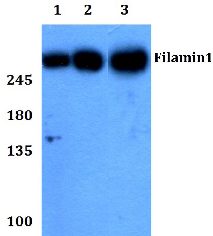
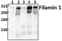
- Western blot analysis of Filamin A in Lane 1: MCF-7 whole cell lysate (20 µg), Lane 2: PC12 whole cell lysate (30 µg), Lane 3: CT-26 whole cell lysate (20 µg). Sample was incubated with Filamin A polyclonal antibody (Product # PA5-86143) using a 1:500 dilution.

- Submitted by
- Invitrogen Antibodies (provider)
- Main image
- Experimental details
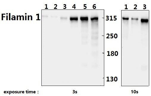
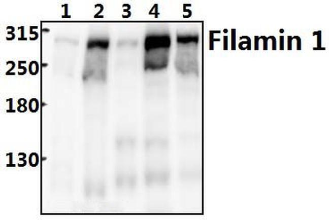
- Western blot analysis of Filamin A in Lane 1: PC12 whole cell lysate (40 µg), Lane 2: CT26 whole cell lysate (40 µg), Lane 3: HEK293T whole cell lysate (40 µg), Lane 4: A549 whole cell lysate (40 µg), Lane 5: MCF-7 whole cell lysate (40 µg). Sample was incubated with Filamin A polyclonal antibody (Product # PA5-86143) using a 1:500 dilution.
Supportive validation
- Submitted by
- Invitrogen Antibodies (provider)
- Main image
- Experimental details
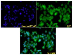
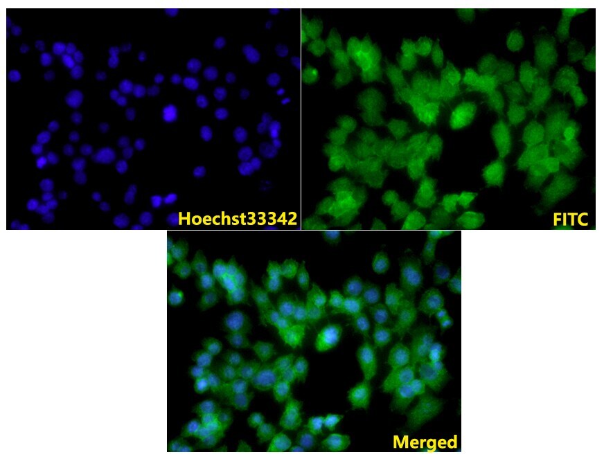
- Immunofluorescence analysis of Filamin A in 3T3-L1 cells. Sample was incubated with polyclonal Filamin A antibody (Product # PA5-86143) using a 1:100 dilution, and followed by goat anti-rabbit IgG (H+L)-FITC and Hoechst (nuclei staining) secondary antibodies at a dilution of 1:200.
- Submitted by
- Invitrogen Antibodies (provider)
- Main image

- Experimental details
- Immunofluorescence analysis of Filamin A in 3T3-L1 cells. Sample was incubated with polyclonal Filamin A antibody (Product # PA5-86143) using a 1:100 dilution, and followed by goat anti-rabbit IgG (H+L)-FITC and Hoechst (nuclei staining) secondary antibodies at a dilution of 1:200.

Supportive validation
- Submitted by
- Invitrogen Antibodies (provider)
- Main image

- Experimental details
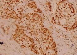
- Immunohistochemical analysis of Filamin A in paraffin-embedded human breast carcinoma tissue. Sample was incubated with polyclonal Filamin A antibody (Product # PA5-86143) using a 1:100 dilution.

- Submitted by
- Invitrogen Antibodies (provider)
- Main image
- Experimental details
- Immunohistochemical analysis of Filamin A in paraffin-embedded human breast carcinoma tissue. Sample was incubated with polyclonal Filamin A antibody (Product # PA5-86143) using a 1:100 dilution.

Supportive validation
- Submitted by
- Invitrogen Antibodies (provider)
- Main image
- Experimental details
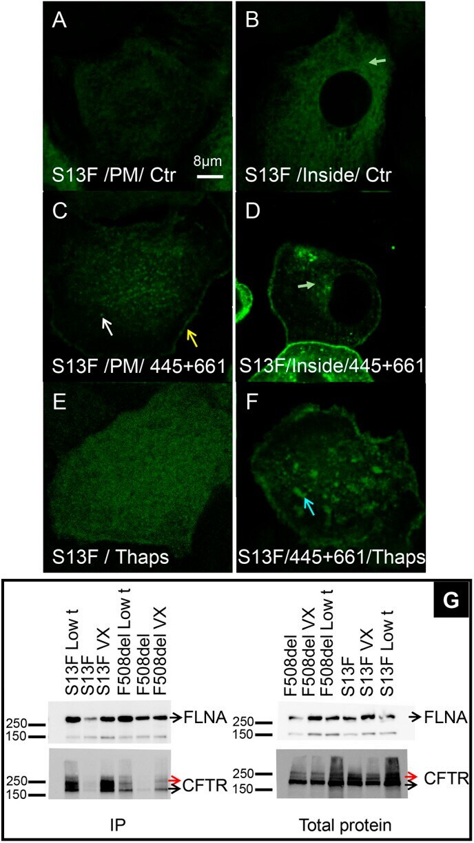
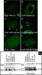
- Fig. 7. Trikafta correctors restore S13F-CFTR folding. HBE cells transduced with adenovirus containing S13F-CFTR. (A) Like F508del-CFTR, S13F-CFTR does not form clusters or a rim near junctions ( N exp =11; n cell =341). (B) Mid-section through cell reveals that most S13F-CFTR is intracellular (green arrow). (C,D) VX-445 plus VX-661 correction reduces S13F-CFTR intracellular retention and restores membrane expression, clustering (white arrow) and rim formation (yellow arrow, N exp =16; n cell =582). The green arrow in D highlights the decrease in intracellular S13F-CFTR level after correction therapy. (E,F) S13F-CFTR fails to form platforms after Thaps treatment ( N exp =3; n cell =90) (E), and Trikafta correctors ameliorate this defect ( N exp =7; n cell =306) (F). The blue arrow in F highlights the restoration of S13F-CFTR incorporation in ceramide-rich platforms after correction therapy. (G) As shown by co-immunoprecipitation, VX-445 plus VX-661 (and low temperature, 29degC for 24 h) restores interaction of S13F-CFTR and F508del-CFTR with FLNA suggesting both mutants are misfolded and rescued by these correctors ( N exp =3). 20 mug of protein were used for the lysate blot and 750 mug for the IP blot (2.7% of the IP).